Playlist
Show Playlist
Hide Playlist
Down Syndrome: Trisomy 21

-
Slides 01 Chromosomal Disorders I.pdf
-
Download Lecture Overview
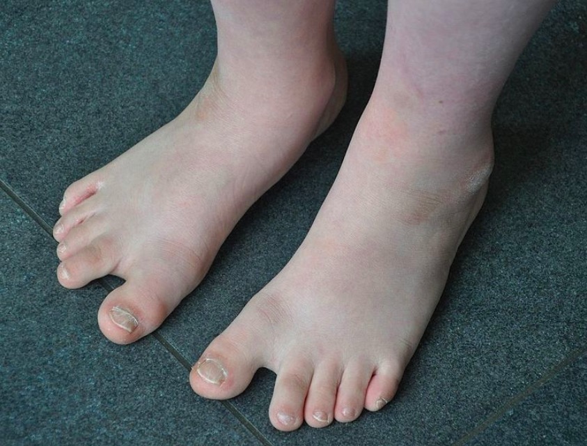
00:01 The neat thing is that only three chromosomal polyploidies actually persist until birth. 00:10 The first of which we’ll consider is Down syndrome. There’s also Edwards syndrome, which is trisomy of 18 and Patau syndrome, which is trisomy of chromosome 13. 00:23 Now, we’re going to spend a lot of time looking at Down syndrome and look also at some visualization techniques or techniques we use in genetics to test for Down syndrome and how those might look. Here, we are with Down syndrome, trisomy 21. You can see in this karyotype clearly. There are three chromosome 21s. Let’s take a look at one of the visualization techniques that we might use during preimplantation or peri-implantation diagnosis or amniocentesis. 01:04 Here is a graphic of FISH, fluorescence in situ hybridization. Here the chromosomes have been stained, 21 has been stained in red fluorescence and chromosome 13 is stained in green fluorescence. 01:22 You can clearly see that there are three chromosome 21s. After amniocentesis sample, the chromosomes can be analyzed. If we see that there’s trisomy of 21, then there are choices that could be made by that mother or parent pair. Here’s another technique that could be used now that we have much more detail in DNA sequencing and microarrays. We examined the technique of microarray and FISH in our previous lectures on molecular genetics and cell biology series. 02:02 Here, you can see clearly that there is an overexpression of the sequences found on chromosome 21 exhibited by the arrow here. So chromosome 21 has extra. We can tell then that there is probably a trisomy of chromosome 21. It may not be the entire chromosome. But we’ll take a look at how that manifests here shortly. Here is another way we might look at it now that the whole genome sequencing is available, again from amniocentesis or preimplantation diagnosis, chorionic villus sampling. 02:39 We’ll look at those techniques later on in the lecture series. But we can see clearly here in this normalized spectrum that chromosome 21 has more gene product than the rest of the chromosomes. 02:54 You can see in the gray area that sort of normal expression. In this purple bar, you can see that there is certainly a higher expression of chromosome 21. Those are three techniques that we could use to diagnose not just the trisomy 21 but any of these chromosomal disorders in which we’re seeing larger chunks of chromosome, either full polyploidies or partial polyploidies. You’ve probably heard that the older a mother is, the more likely we are to see these chromosomal abnormalities. 03:34 Again, that’s because nondisjunction is more likely in older cells with older apparatus. 03:42 In general, we’ll see 1 in about 850 children that are born have Down syndrome. However, you can see by this graphic that it becomes much higher when we look at mothers over 35. We see sort of an exponential increase such that by the time a woman gets to 50 and a fetus gets to a live birth, there’s almost a one in ten chance of the child having Down syndrome which is a very high risk. This is why we should be doing preimplantation diagnosis or amniocentesis and chorionic villus testing if the parents would like to know what the possibilities are or if they do have a conceptus that has one of these disorders. Again, all of those techniques can be used not just for trisomy but many chromosomal disorders. Interestingly, with the systems of checks and balances, our body will spontaneously miscarry most abnormal chromosome situations. Even they say only about 20% to 25% of Down syndrome conceptuses actually make it to birth. The systems in general are working. 05:14 So, it’s not entirely common that we see these disorders, although trisomy 21 is more of the common ones. 05:22 It does persist and survive through sort of middle life into the 30s, perhaps even 40s, even further as our medical systems advance. So, we’ll take a specific look at some of the clinical diagnoses, the features that we might see in a Down syndrome child. They’re really like this nice graphic here of our Down syndrome child. 05:46 But in general, we’ll see a smaller head, smaller face, fairly sparse hair, as well as some changes to the eyes. 05:56 We see that there is fairly short eyelashes as well as bright Brushfield spots, which are small white spots around the iris of the eye. We also might see that the ears are small and fairly lower set, as well as having some abnormalities in the palate. The palate is slightly arched and often we’ll notice a protruding tongue. 06:25 Again, not all of these symptoms will necessarily display but Down syndrome does have a pretty consistent manifestation including this broader, shorter neck. Then, developmental delays are fairly varied in Down syndrome. So, it might be really extreme or it could be fairly light. We’re not entirely sure why that is but again, many conditions are multifactorial. So perhaps environment and nutrition and such have an impact on that. We also noticed that Down syndrome patients have hypotonic muscles meaning that they don’t develop full contraction strength. Then, here is a pretty classic or a couple of classic images of how Down syndrome manifests itself. It’s a pretty characteristic appearance. 07:18 Again, trisomy 21 is one that we should certainly be very familiar with for the USMLE exams because it’s a fairly common trisomy or chromosomal disorder.
About the Lecture
The lecture Down Syndrome: Trisomy 21 by Georgina Cornwall, PhD is from the course Chromosomal Disorders. It contains the following chapters:
- Trisomy 21 - Down Syndrome
- Trisomy 21: Clinical Diagnosis
Included Quiz Questions
Which of the following chromosomal aneuploidies is correctly paired with its name?
- Trisomy 13 - Patau syndrome
- Trisomy 21 - Edwards syndrome
- Trisomy 18 - Down syndrome
- Trisomy 13 - Edwards syndrome
- Trisomy 21 - Patau syndrome
Which of these factors is most strongly linked to a higher incidence of Down syndrome?
- Maternal age
- Paternal age
- Family history of Down syndrome
- Maternal weight
- Paternal weight
Which of the following trisomies is the most common among live births?
- 21
- 11
- 18
- 13
- 22
Which of the following physical characteristics is LEAST associated with Down syndrome?
- Polydactyly
- Protruding tongue
- Brushfield spots
- Short, broad neck
- Flat face
Author of lecture Down Syndrome: Trisomy 21

Georgina Cornwall, PhD
Customer reviews
5,0 of 5 stars
| 5 Stars |
|
5 |
| 4 Stars |
|
0 |
| 3 Stars |
|
0 |
| 2 Stars |
|
0 |
| 1 Star |
|
0 |
