Playlist
Show Playlist
Hide Playlist
Cells: Introduction

-
Slides 04 Types of Tissues Meyer.pdf
-
Download Lecture Overview
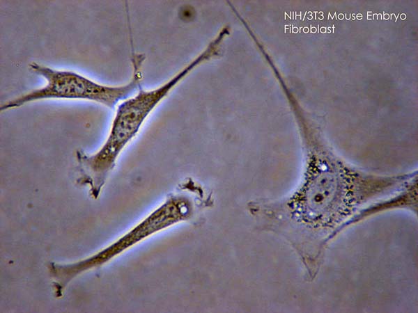
00:01 So let's now move on and look at connective tissue cells. Remember, the three components of connective tissues are fibres, matrix and nerve cells. We will get in more detail and these cells are really derived from mesenchyme. Here is a very very what looks like a complicated diagram with some labels scattered all around. And I put this in this lecture to give you a challenge to make you look at it and try and decide by looking at the left-hand flow of cell differentiation that represents cells that are derived from mesenchyme. 00:48 Look at that derivation of these cells as opposed to those on the right-hand side that are derived from blood. Remember I said early in the lecture, that the cells come under two categories. 01:01 Those that are resident in connective tissue and those that wander in from the blood. 01:07 So my challenge to you, is to have a look at the names of the cells down below and then find their location in one of the two differential pathways, either from mesenchyme or either from blood. And I think it is important that you do that yourself because that helps you understand where each of the cells actually come from and we'll revise through this as we go through this lecture and also as we look at blood later on. 01:39 One of the main connective tissue cells that we should look at, is the fibroblast. 01:45 It makes all the fibres, collagen, reticular fibres and elastic fibres. It also makes the components of the extracellular matrix. Up until now, I have used the term reticular fibre, but really reticular fibres are collagen fibres. They are collagen type III fibres. They are very fine fibres. They were already seen formed the framework of lymph nodes and other hemopoietic tissues that we will talk about later on. The early histologist sort out different sorts of fibres, but later on with more research they are really just found to be normal connective tissue collagen fibres, but of the type 3. So when you hear the term or when you read about the term reticular fibre, just sort of think what they really are collagen fibres, but very fine fibres. Well let's get back to the fibroblast shown here. All you see is a nucleus, sort of rounded up or triangular shaped. It is associated with collagen fibres, you see stained red here. And if you look on the electron micrograph picture, these fibroblasts are full of granular endoplasmic reticulum, that reflects the huge protein factory inside the cell. When you see these fibroblasts actually working, doing their job, during development, you will see details of the cytoplasm. It will be basophilic reflecting the enormous amount of the protein factory within the cell cytoplasm. Here, in fully differentiated adult tissue, the fibroblasts are really just sitting there maintaining the extracellular matrix in the fibres. 03:47 They are not busy, as busy anyway, as what they would have been during early development. 03:53 Sometimes we might refer to these fibroblasts as being resting fibroblasts or fibrocytes. And the top small image shows you the nuclei of one of these fibroblasts and the very extensive cytoplasmic processes. You don't appreciate that when you see each of these sections as the one you see in this main image here. Well let's move on now and have a look at the macrophage.
About the Lecture
The lecture Cells: Introduction by Geoffrey Meyer, PhD is from the course Connective Tissue.
Included Quiz Questions
Reticular fibers are composed of which of the following types of collagen?
- Type 3
- Type 2
- Type 1
- Type 4
- Type 5
Which of the following is NOT synthesized by fibroblasts?
- Triglycerides
- Elastic fibers
- Collagen
- Reticular fibers
- Glycosaminoglycans
Author of lecture Cells: Introduction

Geoffrey Meyer, PhD
Customer reviews
5,0 of 5 stars
| 5 Stars |
|
5 |
| 4 Stars |
|
0 |
| 3 Stars |
|
0 |
| 2 Stars |
|
0 |
| 1 Star |
|
0 |
