Playlist
Show Playlist
Hide Playlist
Hypertrophic Pyloric Stenosis: Examination

-
Slides Hypertrophic pyloric stenosis Special Surgery.pdf
-
Download Lecture Overview
00:01
How do babies present?
Classically, they vomit postprandially.
00:06
The emesis is usually non-bilious.
00:09
And the emesis is described classically as projectile.
00:14
This makes anatomic sense.
00:16
As a pyloric channel becomes stenotic,
gastric content has nowhere to go.
00:21
It’s non-bilious
because it does not have the opportunity
to mix with bilious content in the duodenum.
00:27
Projectile, because
as a stomach gets more and more distended,
it's got a closed channel by the pylorus.
00:33
The only way the patient can drain gastric content
is by forceful vomiting.
00:40
One important thing to notice is how the child is always hungry after vomiting and wants to feed again,
a sign called the hungry vomiter.
00:49
On physical examination,
we oftentimes find an olive mass in the upper quadrant of the abdomen.
00:55
Because babies have relatively thin abdominal walls, this is easier to appreciate.
01:00
On the left side of the screen is a palpation of the abdomen,
finding this olive mass.
01:06
And on the right side of the screen is an ultrasound.
01:09
We’ll get to that in a second.
01:11
Aside from a baby who may have projectile vomiting,
they also have sunken fontanelles.
01:20
What does that mean?
As you’ll remember,
the skull has not completely fused in an infant.
01:25
And one of the first signs of dehydration is sunken fontanelles.
01:29
A lethargic baby.
01:33
This, obviously, is not specific to
hypertrophic pyloric stenosis.
01:37
As you'll recall from many other pediatric modules,
a lethargic baby
or one that has failure to thrive
is an ill baby.
01:45
There are some important findings on chemistry
with patients with non-bilious projectile vomiting,
such as in hypertrophic pyloric stenosis.
01:54
Low sodium,
low chloride,
and low bicarb.
01:59
The CBC may actually be completely normal.
02:02
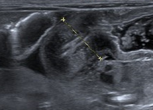
Next, let's move to an ultrasound imaging.
02:06
Ultrasound imaging is first-line therapy.
02:08
Again, it doesn't introduce any radiation to the baby.
02:12
An ultrasound is particularly useful for pyloric channel stenosis.
02:16
Here in this image,
you notice two white dots on the image.
02:20
It’s actually measuring the thickness of the channel.
02:23
Not only is the thickness important,
the length of the channel is also important.
About the Lecture
The lecture Hypertrophic Pyloric Stenosis: Examination by Kevin Pei, MD is from the course Special Surgery.
Included Quiz Questions
How would you describe the vomitous seen in hypertrophic pyloric stenosis?
- Non-bilious
- Bloody
- Bilious
- Pre-prandial
- Non-projectile
Which of the following findings are seen in cases of hypertrophic pyloric stenosis?
- Olive mass in the upper abdomen
- Irregular mass below the right rib cage
- Spongy-feeling mass on both sides of the flank
- Tenderness at McBurney's point
- Tender, irregularly shaped mass in the right upper quadrant
Author of lecture Hypertrophic Pyloric Stenosis: Examination

Kevin Pei, MD
Customer reviews
5,0 of 5 stars
| 5 Stars |
|
1 |
| 4 Stars |
|
0 |
| 3 Stars |
|
0 |
| 2 Stars |
|
0 |
| 1 Star |
|
0 |
Explained in a way to understand very well for all . Presentation is good
