Playlist
Show Playlist
Hide Playlist
Case: 67-year-old Woman with Gait Dysfunction
-
Slides Approach to Dysequilibrium Cerebellum.pdf
-
Download Lecture Overview
00:01
So let's understand
the clinical relevance
of the cerebellar hemispheres,
subcortical white matter
and gray matter
by focusing on a case.
00:09
This is a 67-year-old
African American woman
who presents with a two-year history
of progressive gait dysfunction,
which became progressively worse
after a recent hospitalization
when she was noted
to have severe truncal ataxia.
00:24
The ataxia is improved at rest
and worsens considerably
when she's sitting on the bed.
00:29
She's swaying from side to side.
00:31
Or when she's standing or walking,
so really with any movement.
00:35
Her exam shows
a severe wide base gait,
which we see
with cerebellar problems,
horizontal nystagmus
and saccadic overshoot,
where the eyes are not coordinated.
00:46
when you look in
one direction or the other.
00:48
Finger-to-nose-to-finger test
is slightly abnormal
with mild overshoot,
and again we see or seeing
problems with coordination.
00:56
An MRI is performed and
reveals cerebellar atrophy,
preferentially affecting
the superior vermis
more than other areas
of the cerebellum.
01:05
So which of the following
is the most likely diagnosis?
Well, there are a number of
key features of this case.
01:11
The first is the timeline of onset.
01:13
There's a progressive
two-year history
of this cerebellar disorder.
01:18
That means it's chronic and onset,
and that already us thinking about
some of those acquired causes
of cerebellar dysfunction.
01:26
The second is the
propagating factors.
01:28
This patient's dizziness
or balance problem
is provoked by moving
and improved with rest.
01:34
That's very common
with cerebellar pathology.
01:37
It can be seen with vertigo
and vestibular pathology.
01:40
But that movement component is
something that the cerebellum does.
01:43
So when we see problems
that are provoked by movement,
we want to think
about the cerebellum.
01:49
In addition,
this patient's exam
shows problems with
cerebellar dysfunction,
wide base gait, nystagmus,
saccadic overshoot,
and difficulty with
finger nose finger,
are all cerebellar signs.
02:00
That means
this dizziness problem
is a problem with the
cerebellum and disequilibrium.
02:06
Let's look more closely
at this patient's MRI scan.
02:10
We see atrophy of the cerebellum,
and here we're looking
at a midline cut.
02:15
This is a sagittal MRI,
looking right at the middle
of the cerebellum,
right at the area of the vermis.
02:21
We see atrophy across
the cerebellum,
but preferentially affecting the
upper part of the cerebellum,
that superior lobe,
that top lobe along the vermis.
02:31
And there are certain conditions
that will affect
only the hemispheres,
only the vermis,
and one that we think about
that has a predilection
for the superior cerebellar vermis.
02:42
So what's the most likely
diagnosis for this patient?
Is this a post-infectious
cerebellitis,
a Chiari malformation,
alcohol-related cerebellar ataxia,
or a cerebellar stroke?
Well, this doesn't sound
like a post-infectious cerebellitis.
02:57
Typically, we would recognize
the initial infection,
which we don't see in this case.
03:01
Post-infectious cerebellitis
is an inflammatory condition.
03:05
It's often subacute and onset,
and this is chronic and onset.
03:10
It's been going on
over multiple years,
or two years for this patient.
03:14
In addition,
and most importantly,
post-infectious cerebellitis
typically affects the hemispheres.
03:19
And this is a problem
that is very specific
to the cerebellar vermis.
03:23
So post-infectious cerebellitis
is not the most likely diagnosis
in this patient.
03:30
Cerebellar stroke is also unlikely
for this patient.
03:33
Strokes present acutely.
03:35
And again, this patient's symptoms
was chronic and onset
over two years.
03:39
In addition,
we often see strokes
affecting the lateral
circumferential vessels
that feed
the cerebellar hemispheres,
and would present with prominent
appendicular dysmetria and ataxia
as opposed to vermis dysfunction.
03:56
Chiari malformation
often presents with headache
and really uncommonly presents
with an ataxic syndrome.
04:02
A Chiari is a description
of malformation
for the cerebellar tonsils
descending down
below the foramen magnum.
04:10
We don't see that on imaging
on that midline cut,
and it doesn't have
this type of presentation.
04:16
And so this patient
is suffering from
alcohol-related cerebellar ataxia.
04:20
The patient has
an ataxic syndrome
with a predilection
or that superior vermis,
which is something
that's very typical
for alcohol-related
cerebellar syndromes.
04:31
So let's talk
a little bit more about
Cerebellar Vermis Atrophy
in Alcoholism.
04:35
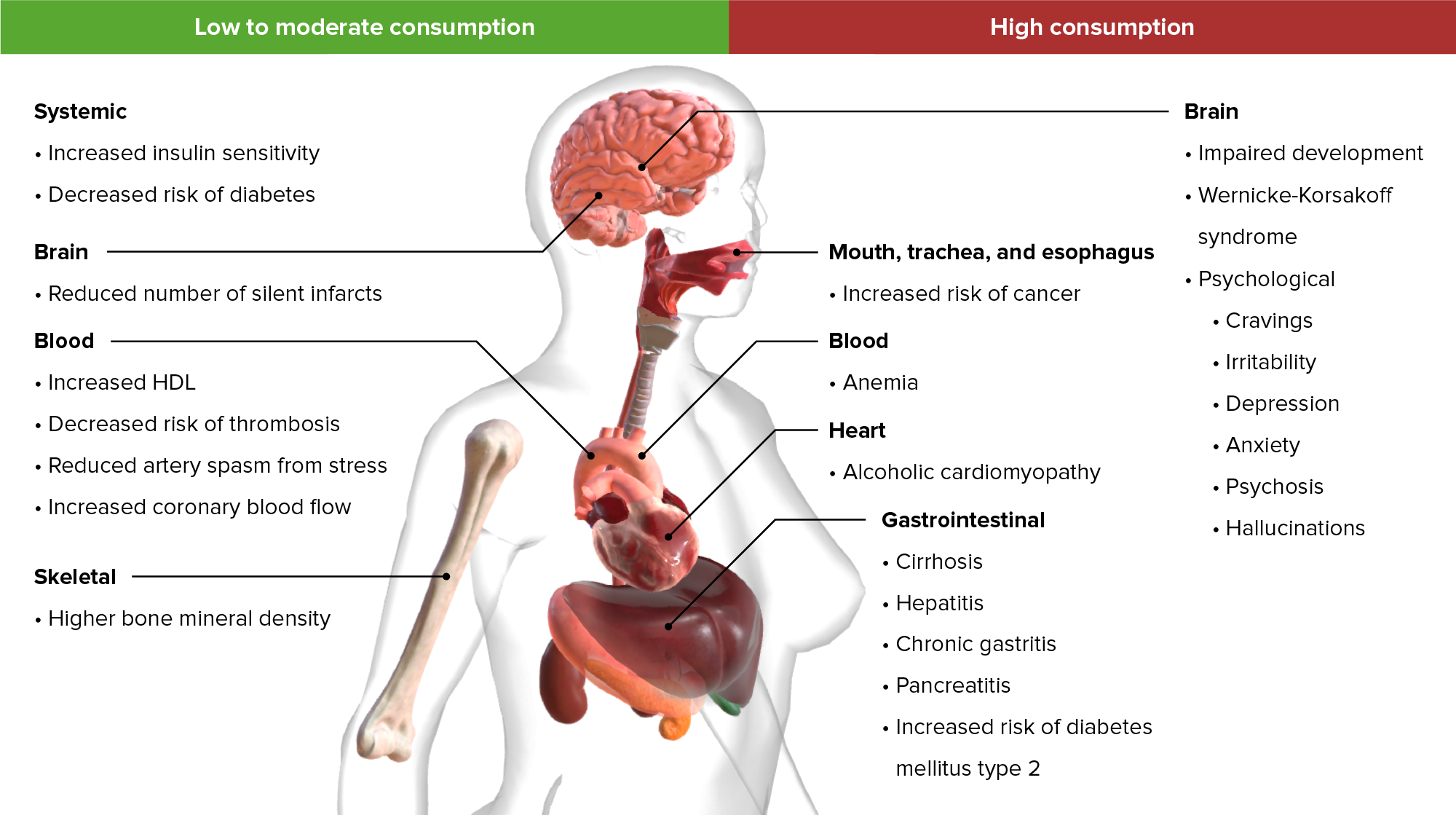
This is the clinical relevance
of understanding,
how the cerebellum is organized?
In terms of definition,
this is a primary long-term effect
of chronic alcoholism,
which results in degeneration
or atrophy of the cerebellum.
04:49
The entire cerebellum is involved
and alcohol has a predilection
for affecting the
cerebellar purkinje fibers,
but the superior vermis
is most specifically affected,
and often one of the
earliest areas that we see.
05:02
Patients typically present with a
slowly progressive gait dysfunction
and truncal ataxia.
05:07
The vermis is involved
in truncal coordination.
05:10
And that's more so affected
than the appendicular functions,
as in this case.
05:15
On imaging, MRI shows atrophy of
the diffusely across the cerebellum,
but preferentially affecting
the superior cerebellar vermis
more so than the hemispheres.
05:25
And pathologically,
what we see
is loss of cerebellar
and purkinje cells
primarily
in the superior vermis,
as well as the vestibular nuclei.
05:33
And so we can see,
eye movement dysfunction
as was present in this case,
and can be seen
in up to 43% of patients.
05:39
So it's common,
something we look for.
About the Lecture
The lecture Case: 67-year-old Woman with Gait Dysfunction by Roy Strowd, MD is from the course Vertigo, Dizziness, and Disorders of Balance.
Included Quiz Questions
Which of the following would suggest cerebellar pathology?
- Nystagmus
- Vertigo
- Unequally dilated pupils
- Bilateral leg weakness
- Narrow gait
Which region of the cerebellum is most commonly impacted by a stroke?
- Lateral hemispheres
- Subcortical gray matter
- Flocculonodular lobe
- Inferior vermis
- Superior vermis
Which statement is the most accurate with respect to alcohol-related cerebellar ataxia?
- The superior vermis is highly susceptible to alcohol toxicity.
- Patients show an acute onset of symptoms.
- Symptoms are isolated to the appendages.
- MRI usually shows isolated degeneration of the lateral hemispheres.
- The lateral hemispheres are not susceptible to alcohol toxicity.
Customer reviews
5,0 of 5 stars
| 5 Stars |
|
5 |
| 4 Stars |
|
0 |
| 3 Stars |
|
0 |
| 2 Stars |
|
0 |
| 1 Star |
|
0 |


