Playlist
Show Playlist
Hide Playlist
Hemorrhagic Shock: Locate Blood & Perform E-FAST Exam


-
Emergency Medicine Abdominal Injuries and Hemorrhagic Shock.pdf
-
Download Lecture Overview
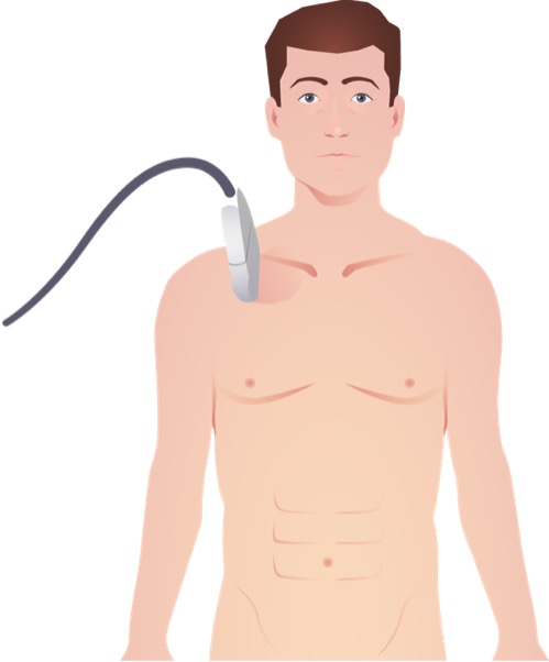
00:01 Alright, now when we think about hemorrhagic shock, one of the questions we always have to ask ourselves is where is the blood? You need to identify where the blood is in the body and where it’s gone, so that you can plan your treatment accordingly. 00:17 So blood is not gonna be in the head if your patient is in hemorrhagic shock except maybe in infants, right? The head in adults is not large enough to hold enough blood volume to cause shock. 00:29 Now, infants have relatively larger heads compared to their body sizes and their heads are distensible. 00:34 So there actually are cases where infants with large intracerebral hemorrhages can be in shock. 00:40 But typically, we’re not gonna think about the head. 00:42 What we are gonna think about is the thorax. 00:45 Each hemithorax can hold a liter to a liter and a half of blood, so that’s definitely enough to put you at least in the early stages of shock. 00:53 Blood’s not gonna be in the extremities. 00:55 They typically do not hold enough blood volume for that. 00:58 You can maybe lose a unit of blood into each thigh from a femur fracture. 01:03 But again, that’s not really gonna be enough to cause significant shock. 01:07 The abdomen however, definitely can hold enough blood volume for that. 01:11 The abdomen is large, it’s readily distensible, it’s very easy to put a lot of blood into it. 01:16 So interabdominal injuries are commonly associated with shock. 01:20 As our pelvic injuries for the same reason. 01:23 But the neck, definitely not an area where you’re gonna be able to get hemorrhagic shock unless you’re exsanguinating externally, and that’s the caveat with all of these. 01:34 The other places the blood can be in every case is on the floor, at the scene, etc. 01:40 So remember, patients bleed internally but they obviously also bleed externally. 01:46 So you always wanna get a sense from your paramedics about how much blood was at the scene, how much blood was lost in the ambulance, and you wanna get a feel for how much ongoing external blood loss there isin the ED to see if that could potentially account for the hemorrhagic shock that you’re seeing in your patient. 02:02 You have identify where the blood has gone because this is gonna dictate how you manage the patient. 02:07 A patient with intraabdominal hemorrhage needs operative management. 02:10 A patient with thoracic hemorrhage needs chest tubes, etc. 02:14 So you need to understand where the blood is so that you can treat the patient appropriately. 02:20 So that pretty much covers hemorrhagic shock. 02:23 The other type of shock to be aware of in trauma is obstructive shock which is caused by injuries like cardiac tamponade and tension pneumothorax. 02:31 Obstructive shock occurs when there is an extra cardiac obstruction to normal blood flow. 02:37 In the case of both of these disease processes, there’s actually mechanical compression of the heart. 02:42 In the case of pneumothorax by an air collection. 02:45 In the case of cardiac tamponade by a pericardial fluid collection or blood collection. 02:51 In both cases, the compression of the heart mechanically impedes venous filling. 02:57 And of course, if the heart doesn’t fill normally during diastole, it can’t pump blood out to the body normally which is gonna lead to shock. 03:05 We’ve addressed tension pneumothorax in another lecture and we’re gonna talk about cardiac tamponade at the end. 03:11 So what diagnostic test can identify all the potential etiologies for shock in trauma patients? Wouldn’t it be great if there was just one thing we could do to really get to the bottom of why our patient is in shock? Well, it turns out that there is and that’s the E-FAST which is the extended focused abdominal sonography in trauma exam. 03:33 And it’s basically a bedside ultrasound exam that’s designed to localize where the blood is if the patient is in hemorrhagic shock. 03:41 It’s designed to identify hemopericardium and pneumothorax, so that we can address our causes of obstructive shock. 03:48 It could be performed right there at the bedside as part of the secondary survey, and it was originally developed for use in unstable blunt trauma patients. 03:56 But it actually can be used in any trauma patient as an adjunct to your physical exam to give you a better sense of what’s going on with the patient. 04:05 So there are five views that we do as part of the E-FAST exam. 04:08 There’s the right upper quadrant, the left upper quadrant, the bladder view, the pericardial view, and the bilateral lung views. 04:23 The bottom line on E-FAST is that you’re answering three yes/no questions and those questions are, is there hemorperitoneum? Is there hemopericardium? And is there pneumothorax? What you’re not answering with E-FAST is, what is the injured organ? How much blood is in there? How badly is it injured? Is it going to be treatable conservatively or is it going to need surgery? You’re not answering those questions. 04:52 You are simply answering questions about whether there is unauthorized blood or air in major body cavities and that’s really important to remember. 05:03 So when we think about ultrasound examinations, a few basic principles will help us interpret our images. 05:10 Fluid on ultrasound is anechoic, it’s always gonna appear as black on your image. 05:16 So when you look at blood vessels, when you look at the chambers of the heart, they’re all gonna be filled with black. 05:22 Air on the other hand is hyperechoic. 05:25 Air appears as white on ultrasound and then tissue based on its consistency and composition is gonna be varying shades of grey in between. 05:34 The goal of the E-FAST again is to detect blood or air in places where they don’t belong. 05:39 So our goal as we perform this exam is gonna be to look for black or white in places where we don’t want to see it.
About the Lecture
The lecture Hemorrhagic Shock: Locate Blood & Perform E-FAST Exam by Julianna Jung, MD, FACEP is from the course Trauma (Emergency Medicine).
Included Quiz Questions
What diagnostic test can identify all potential etiologies of shock in trauma patients?
- eFAST
- CT scan
- MRI scan
- Radiograph
- Angiogram
What is the color of fluid in an ultrasound evaluation?
- Black
- White
- Gray
- Green
- Red
Which of the following statements regarding eFAST is INCORRECT?
- It provides information regarding the severity of the injury.
- It does not provide information on the location of the injury.
- It can detect presence of hemoperitoneum.
- It can detect presence of pneumothorax.
- It can detect presence of hemopericardium.
Author of lecture Hemorrhagic Shock: Locate Blood & Perform E-FAST Exam

Julianna Jung, MD, FACEP
Customer reviews
5,0 of 5 stars
| 5 Stars |
|
5 |
| 4 Stars |
|
0 |
| 3 Stars |
|
0 |
| 2 Stars |
|
0 |
| 1 Star |
|
0 |
