Playlist
Show Playlist
Hide Playlist
Pupillary Light Reflex


-
Slides 15 VisualPathway BrainAndNervousSystem.pdf
-
Download Lecture Overview
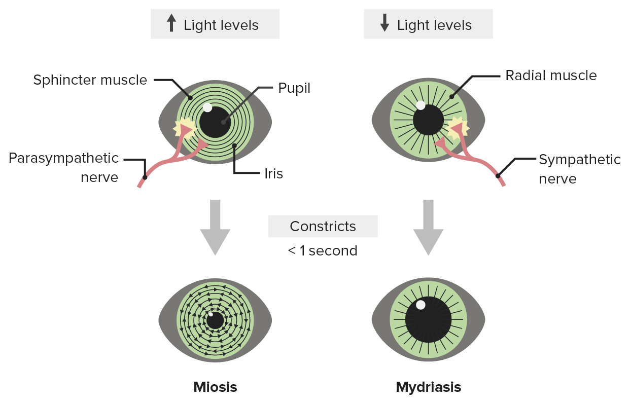
00:00 Next, I want to guide you through the pupillary light reflex. So in this visual, we have two eyeballs. This happens to be the one on the left. We also have an eyeball here. 00:19 That would be the right eyeball of the person looking at you. We are now going to shine light in the left eye. So we’ll shine the light in through here. The light will travel through the structures, components of the left eyeball, hit the retina. Then ultimately, ganglion retinal cells will be stimulated. Then the action potentials will travel through the optic nerve to the pretectal region of the brain. It’s going to be on the left side here of that area. 01:00 Then this will activate nerve cells that will communicate bilaterally to the Edinger-Westphal nuclei. So we see in this simple illustration, we have a nerve cell sending an axon here to activate the left Edinger-Westphal nucleus. Then the contralateral or the right one is also going to be stimulated in response to this pupillary light reflex. From the Edinger–Westphal nuclei, what will happen here is there will be parasympathetic fibers that will leave this nucleus. They’re going to travel to the ciliary ganglia on both sides. So here’s the left one to the left eye, ciliary ganglion then to the right eye. These parasympathetic fibers are going to travel in the oculomotor nerve as we see identified here. From the ciliary ganglia, nerve fibers are going to innervate the sphincter pupillae muscle of each eye and cause it to contract. So what we have here would be direct and consensual light reflexes. 02:25 The direct reflex is that the light shined in the left eye will cause constriction. The consensual reciprocal response is that the right eye will do the same in response to that light stimulus. 02:41 Accommodation is another mechanism that’s associated with the eye. Accommodation is necessary to provide focus upon objects that we are viewing close to us. For near vision, the structures that we have here would be the Edinger-Westphal nucleus, which we saw as a principal player in the pupillary light reflex. We also have our oculomotor nerve again and our ciliary ganglia. So what we’ll have is preganglionic parasympathetic fibers will travel from the Edinger-Westphal nucleus via the oculomotor nerve. They will then synapse within the ciliary ganglion. Then from here, postganglionic parasympathetic fibers will innervate the ciliary muscle, which is shown in this area here and on the opposite side here. 03:50 The lens is in the center here. As a result of that innervation, the ciliary muscle will be prompted to contract. This will decrease the tension on the suspensory ligaments that are maintaining the thinness of the lens at this point. As the suspensory ligaments lose their tension, the lens will increase its thickness or convexity, as we see here. That will more sharply bend the light to focus it on the fovea centralis when we’re looking at objects that are close to us. This would be a bilateral or reciprocal reflex, so it would be occurring in both eyes.
About the Lecture
The lecture Pupillary Light Reflex by Craig Canby, PhD is from the course Visual Pathways. It contains the following chapters:
- Pupillary Light Reflex
- Accommodation
Included Quiz Questions
Which cranial nerve is connected to the Edinger-Westphal nucleus?
- CN III
- CN V
- CN I
- CN VII
- CN X
Which of the following structures does oculomotor CN III innervate in regard to the pupillary light reflex?
- Sphincter pupillae muscle
- Ciliary muscle
- Macula lutea
- Lens
- Suspensory ligament
During the accommodation reflex, which of the following structures does oculomotor CN III innervate?
- Ciliary muscle
- Suspensory ligament
- Macula lutea
- Lens
- Sphincter pupillae muscle
Author of lecture Pupillary Light Reflex

Craig Canby, PhD
Customer reviews
5,0 of 5 stars
| 5 Stars |
|
5 |
| 4 Stars |
|
0 |
| 3 Stars |
|
0 |
| 2 Stars |
|
0 |
| 1 Star |
|
0 |
