Playlist
Show Playlist
Hide Playlist
Abdominal Radiography

-
Slides Abdominal Radiography Technique.pdf
-
Download Lecture Overview
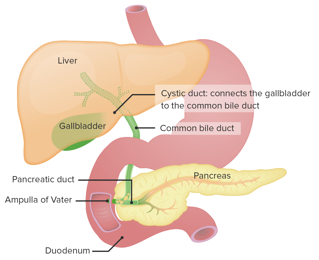
00:01 So before we delve in to the many abnormalities that can be found within the abdominal x-ray, let's review first some of the normal findings on an abdominal film on a normal approach. 00:11 So most abdominal imaging and diagnosis involves the use of ultrasound, CT, or MRI. 00:18 However, radiographs are often used as a quick first step to help you decide what further imaging is needed next if any. 00:25 So what can be evaluated on abdominal plain films? The bowel gas pattern is a very important thing. 00:31 You can always evaluate for free air which is something that you really don't wanna miss. 00:35 Abnormal calcifications can be seen on abdominal radiographs or abnormal masses. So these are the 4 major points that you wanna keep in mind when you're taking a look at an abdominal radiograph. 00:45 So let's come up with a general approach. 00:49 As with all of radiology, it's important to have a standard approach so that you make sure that you look at every aspect of the film. 00:54 So what I do is I start off with the lung bases, even though we're performing an abdominal film often times a patient may have an abnormality at the lung base that you don't wanna miss I then look for free air because this is a finding that you really don't wanna miss on an abdominal plain film. Next, I look at the bowel gas pattern and then I evaluate the solid organs. 01:13 I then look for any abnormal soft tissue masses, any abnormal calcifications, and lastly I take a look at all of the bony structures. 01:21 So before we look at the abdominal normal anatomy on a radiograph, let's just review what normal abdominal anatomy looks like on a diagram. 01:31 So a large part of the upper abdomen is encompassed by the liver. 01:35 It's essentially seen on the right upper abdomen but it also extends over to the left upper abdomen. 01:40 Adjacent to it is the spleen and often times on a radiograph you can see the outline of both the spleen and the liver. 01:47 The gallbladder is usually not well seen on an abdominal plain film, however, if it has calcifications in it such as gallstones that may be visualized. 01:55 And then the majority of the rest of the abdomen is encompassed by large bowel which is seen peripherally and then small bowel which is usually seen centrally. 02:04 So let's review some radiographic anatomy. 02:09 So again, I start off by taking a look at the lung bases and you can see them on every abdominal radiograph. 02:15 So you can see the diaphragm here on the right and then you can see the left hemidiaphragm here and just above it are the lung bases and a small portion of the heart. 02:24 In the left upper abdomen you can see the stomach bubble. 02:29 It usually has a little bit of air and then it has an air fluid level which is, again, normal. You can see the outline of the liver, primarily encompassing the right upper abdomen and then you can see air within the large and small bowel. 02:42 In terms of bony structures, you can take a look at the iliac crest which really should be included on every abdominal radiograph and you can see the thoracic and lumbar spines. 02:53 So how can you differentiate between small and large on a radiograph? When there's a bowel abnormality it's important to try and see which bowel it's involving. So the small bowel is located centrally and it has valvular markings called valvulae conniventes. 03:10 They cross the entire width of the small bowel and they're spaced very close together. 03:14 They have the classic stacked coin appearance so if you take a look at these stacks of coins the small bowel actually looks very similar to that and that's a very classic appearance of small bowel. 03:25 Large bowel on the other hand is usually located peripherally and it contains haustra. 03:31 So the haustra either do not connect from one wall to the other or they're spaced a lot wider apart than the valvulae conniventes are. 03:39 So if you take a look at this image here, the haustra do actually go all the way from one end of the wall to the other. 03:44 However, they're not the typical stacked coin appearance that you would expect from a small bowel, they are much wider apart. 03:51 This is another example of the large bowel again seen predominantly in the periphery of the abdomen and you can see air within small pieces of small bowel seen more centrally. 04:05 So how do you assess bowel gas pattern? A normal bowel gas pattern in the small bowel usually has about 2 or 3 air fluid levels. 04:15 It's normal to see air within the small bowel and usually the bowel is not more than about 3 cm in diameter. 04:21 Large bowel on the other hand usually doesn't have air fluid levels because a lot of the fluid is absorbed within the large bowel. 04:28 It does contain air and that's normal and it can have a very variable diameter so it's hard to rely on large bowel diameter to tell whether or not it's abnormal. 04:37 So let's take a look at this, this is the coned down view of the right upper abdomen. 04:43 You can see a somewhat modeled appearance here. 04:46 So what do you think that represents? It doesn't follow the typical features of small or large bowel. 04:51 So this is an example of normal stool within the colon. 04:55 It appears as very small bubbles of gas that are partially solid and they appear within the expected location of the colon. 05:02 So abdominal films can be performed in 3 different projections and each one has different uses. 05:12 So typically, it's performed as an upright film. 05:14 Air is gravity dependent so it's very useful to evaluate for free air so the air will rise up to the top and can often be seen just underneath the diaphragm. 05:24 It's also used to evaluate for air fluid levels which can indicate stasis and ileus or bowel obstruction. 05:30 So when the patient is standing upright, again, the air will rise to the top of the bowel and you can see the air fluid level because the fluid will be at the bottom of the bowel. 05:38 Usually, these are obtained with the patient sitting or ideally in the standing position. 05:43 A left lateral decubitus is usually used in patients that aren't able to stand upright. 05:49 So this is performed really as just a substitute for an upright film. 05:52 It also allows for evaluation of air fluid levels and it allows for evaluation of free air so the patient lies on their left side down, the right side is up and again air will rise to the top part of the body and so you can see it adjacent to the liver on the right here. 06:11 Ideally, again it's performed on the left side down because the liver forms a solid organ on the right which allows you to see the difference between the air and the density of the liver. If you perform it the other way, you have a lot of bowel on the left side and you may not see the difference between air within the bowel and air outside of the bowel. 06:29 So supine film is also very commonly performed and it's usually performed in addition to an upright or a decubitus. 06:37 This provides a general overview of the abdomen and it's the one that's used to look for calcifications or abnormal soft tissue masses. 06:45 This is obtained with the patient lying on their back and it's really best when it's used again in conjunction with another examination, either the upright or the decubitus. 06:54 So incidentally, this patient has a finding in their pelvis. 06:58 So what do you think this is? This is actually an intrauterine device or an IUD. 07:10 It's used as a contraceptive device and this is very commonly seen in women. 07:14 This is a T-shape metallic structure which is why it appears so dense. 07:19 It's actually a little bit more dense than the surrounding bony structures. 07:22 So the take away points from this lecture I think are that you really need to look for air fluid levels and free air within the abdomen. 07:30 Those are the 2 most important findings and these are really best seen on an upright or a decubitus view. 07:36 Free air is mostly easily seen on these views as well and it's a finding that you really don't wanna miss. 07:41 So hopefully, now that you have the background knowledge of the abdominal anatomy, we can move on to discussing some pathology and the appearance of pathology on an abdominal plain film.
About the Lecture
The lecture Abdominal Radiography by Hetal Verma, MD is from the course Abdominal Radiology. It contains the following chapters:
- Abdominal Radiography: Normal Findings & Approach
- Upright Abdomen, Left Lateral Decubitus, Supine Abdome
Included Quiz Questions
Which statement regarding small and large bowel is CORRECT?
- The small bowel generally contains 2-3 air-fluid levels.
- The small bowel is variable in diameter.
- The large bowel should not be more than 3 cm in diameter.
- The small bowel should not usually contain air.
- The large bowel has valvulae conniventes.
A 54-year-old male is brought to the emergency room with complaints of abdominal pain of gradual onset. He has a long history of hypertension and gastric ulcer. An abdominal radiograph is usually NOT used to identify which among the following?
- Free fluid
- Free air
- Bowel gas pattern
- Abnormal calcifications
- Abnormal masses
Which organ gives the classic stacked coin appearance on an abdominal radiograph?
- Jejunum
- Ascending colon
- Transverse colon
- Rectum
- Esophagus
Which of the following regarding the left lateral decubitus position for an abdominal X-ray is FALSE?
- It is usually used for calcifications and soft tissue masses.
- It is performed as a substitute for an upright film in patients who have had a recent hip replacement surgery.
- It allows the evaluation of air-fluid levels.
- It allows visualization of free air around the liver.
- It is performed with the patient lying on the left side.
Author of lecture Abdominal Radiography

Hetal Verma, MD
Customer reviews
5,0 of 5 stars
| 5 Stars |
|
1 |
| 4 Stars |
|
0 |
| 3 Stars |
|
0 |
| 2 Stars |
|
0 |
| 1 Star |
|
0 |
Concise and well presented. Delivered all the necessary information without wasting any time
