Playlist
Show Playlist
Hide Playlist
How are Viruses Studied?

-
Slides 04 Viruses MicrobiologyAdvanced.pdf
-
Download Lecture Overview
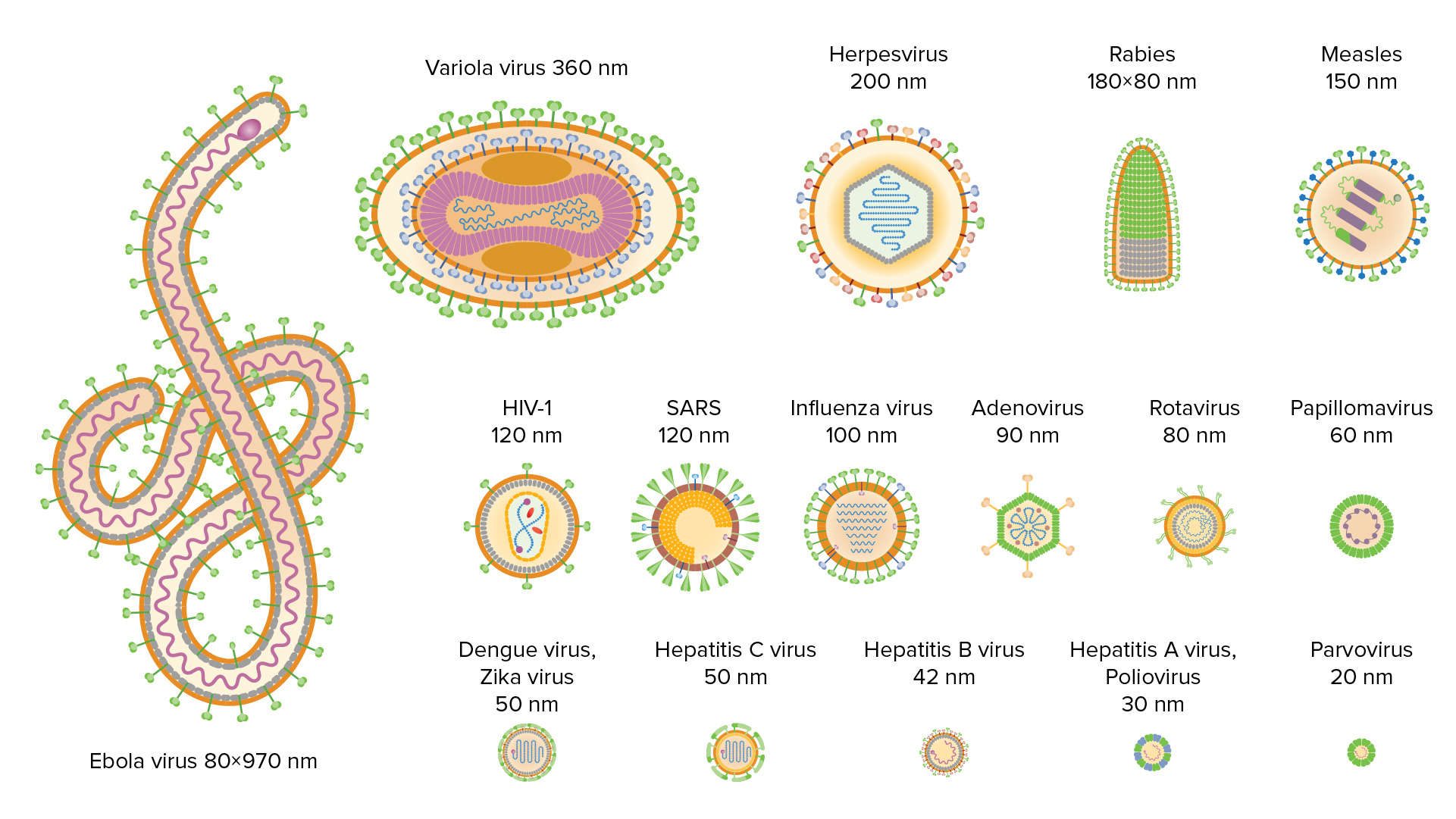
00:01 Hello and welcome to viruses. 00:04 We're going to dig a little bit deeper into this fascinating subject, and at the end of today's lectures, you should be able to know how viruses are cultivated and assayed. 00:16 You should be able to distinguish the seven different types of viral genomes and their structures. 00:23 And you should appreciate what information is and is not encoded in a viral genome. 00:32 Let's first start by talking a little bit about how we study viruses. Now you remember from our introductory lecture that animal viruses were discovered around the end of the eighteen hundreds, but for many years, these viruses could not be routinely studied in cells in culture. 00:54 They had to be studied, and laboratory animals and laboratory animals of all sorts were used. 00:59 Some of them are shown here. 01:01 But as you can imagine, this is not a convenient procedure. 01:05 Plus, laboratory animals can differ from experiment to experiment. 01:10 So it was very important to try and determine how to grow viruses in cells, and many scientists worked on that problem before nineteen forty nine. It was not possible to use cells in a consistent and reproducible way to study viruses. 01:27 But in that year, three scientists, Enders Weller and Robbins, working in the U.S., found that they could propagate polio virus, a human virus, in human cell cultures. 01:41 Now they happen to use what we call primary cultures from embryonic tissues. 01:46 But this experiment set a precedent for the first time it was possible to propagate a human virus in cell cultures. 01:53 This was a very important discovery for which these researchers receive the Nobel Prize in Medicine in nineteen fifty four. 02:03 That discovery is with us today. 02:06 We since nineteen forty nine, we've been able to study viruses in a variety of cell cultures derived from different sources. 02:15 For example, we can use primary cell cultures where the tissue is taken from a source it's minced up to to produce individual cells, and then it's put on a plastic layer, in addition covered with medium. 02:31 So on the left hand part of this slide, you can see a photograph of primary human foreskin fibroblast cells in culture. 02:40 Human for skins are very easy to obtain because when babies are born, we often remove the foreskin. 02:47 It's thrown away in the hospital. 02:49 But if you're interested, you can obtain them and produce primary cells like these. 02:53 This, of course, is not so convenient to use on a routine basis. 02:57 So for that reason, we have what we call cell lines. 02:59 And there are two examples of of these cell lines shown on this slide. 03:03 One is a mouse fibroblast cell line. 03:06 It's called three T three, and then there is a human epithelial cell line. The famous HeLa cells, both of these cells are what we call immortal. They grow forever. 03:18 You can grow them over and over in the laboratory, and then that makes it a lot easier to do your experiments. 03:25 Now, the negative aspect of using a cell line is that they're a little bit unusual. 03:30 They have far too many chromosomes, they're a bit abnormal, and some of them are, in fact, cancer cells. 03:36 So if this is a problem for your research, we also have what we call diploid cell lines like the W.I thirty eight cell line, which is derived from human embryonic lung. These have a normal number of chromosomes. 03:49 They don't live forever, but they do live a long enough time to make your experiments convenient.
About the Lecture
The lecture How are Viruses Studied? by Vincent Racaniello, PhD is from the course Viruses.
Included Quiz Questions
Which of the following statements about viruses is true?
- Viruses can grow in cell culture.
- Viruses cannot replicate without the Golgi apparatus.
- Viruses are autotrophs.
- The viral envelope does not contain any protein.
- The viral capsid is made up of carbohydrates.
Author of lecture How are Viruses Studied?

Vincent Racaniello, PhD
Customer reviews
2,0 of 5 stars
| 5 Stars |
|
0 |
| 4 Stars |
|
0 |
| 3 Stars |
|
0 |
| 2 Stars |
|
1 |
| 1 Star |
|
0 |
not detailed and for a introductory vedio should be more explanatory
