Playlist
Show Playlist
Hide Playlist
Knee Joint – Joints of Lower Limb
-
Slides 09 LowerLimbAnatomy Pickering.pdf
-
Download Lecture Overview
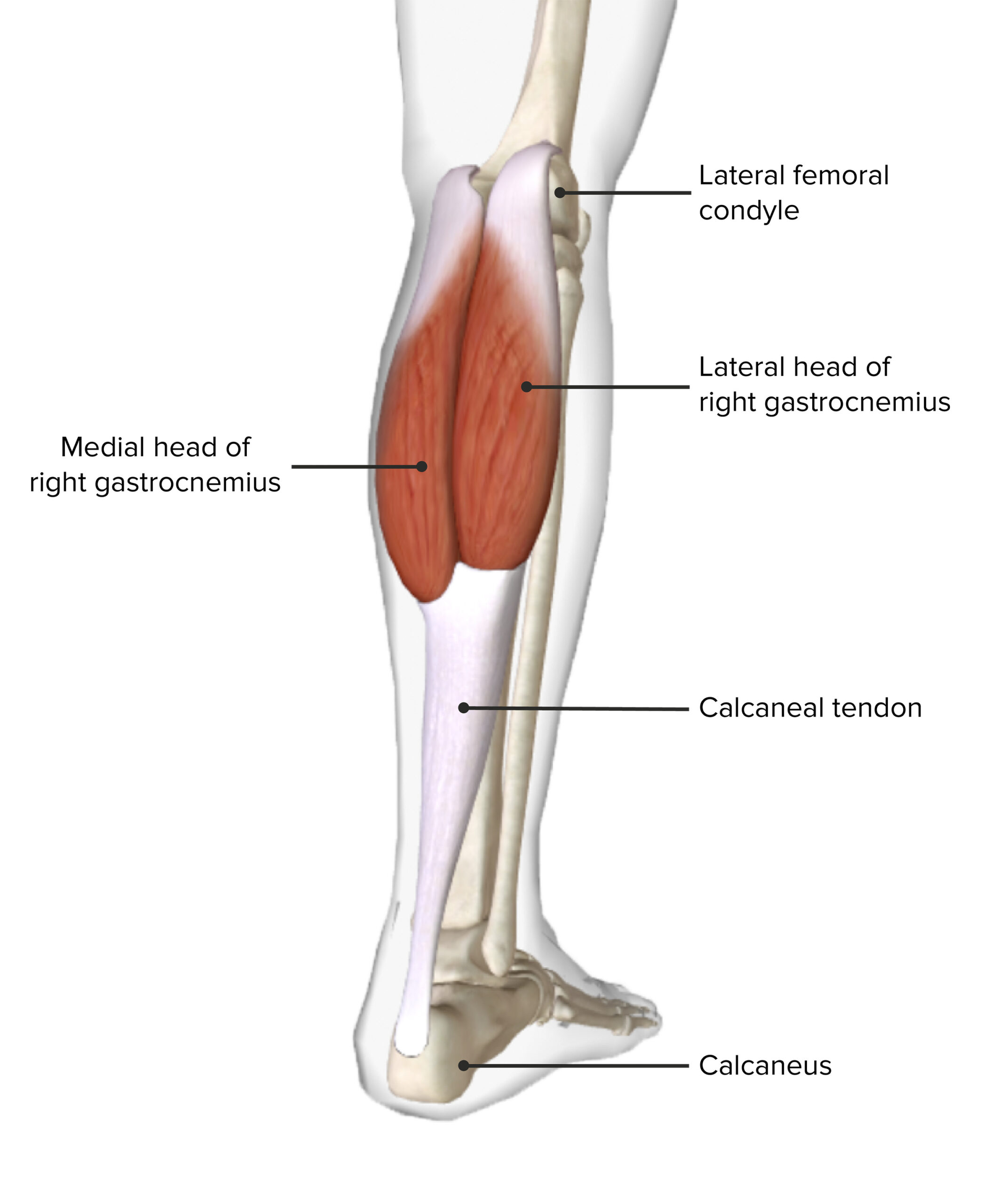
00:00 Now let’s move on to the knee joint. And we can see here both the bony articulations of the knee joint on this side, and we can see the various joint capsule and ligaments of the knee joint over here. So we can see there are three articulations with the knee joint. We have the femorotibial articulations where there were two between the lateral and medial femoral condyles and the tibial condyles. So we can see here the lateral condyle of the femur articulating with the tibial condyle of the tibia, the lateral tibial condyle. And here we can see the articulations between the medial femoral condyle and the medial tibial condyle. 00:43 We also have the femoropatellar articulation. And that is between the patella and the distal surface of the femur on its anterior surface. The joint capsule superiorly is the margin of the articular surface of the femoral condyles. So the joint capsule is superior to where the articular surface stops. Inferiorly, the margin of the articular surface is on the tibial plateau although it does have an opening for the popliteus muscle. So the joint capsule surrounds the knee joint and it surrounds the articular surfaces. Internally, the joint capsule is lined by this synovial membrane consistent with synovial joint capsules, and except where they invaginate to surround the cruciate ligaments. So the synovial membrane lines all of the joint capsule, but at some point, is reflected posteriorly and they invaginate around the cruciate ligaments. This keeps the cruciate ligaments extrinsic, keeps them outside of the synovial cavity. It also subdivides the articular cavity into right and left half. 01:57 So we have the invagination as we’ll see of the synovial membrane posteriorly. 02:04 Let’s have a look at the joint capsule and the various ligaments of the knee joints. Here, we can see both an anterior aspect of the knee joint with the muscle still attached forming the patellar tendon or the patellar ligament from quadriceps muscle. And here we can see it opened up to see the patellar surface and the articular patellar surface of the femur. 02:26 Here we can see the various ligaments and the fibrous capsule that surrounds the knee joint where it’s been opened up here to look into the synovial cavity. So the joint capsule is going to be strengthened by series of intrinsic ligaments. There’s the patellar ligament which we can see most anteriorly, running from the patella down to the tibial tuberosity. And this is the distal part of the quadriceps tendon. It receives the medial and lateral retinacular from the medial and lateral vasti. So we can see coming into the patella here and passing down towards the tibia is the medial patellar retinaculum and the lateral patellar retinaculum. And these are passing down from those muscles to help reinforce the anterior aspects alongside the patellar ligament of the knee joint. We also have the oblique popliteal ligament, and this is a reflected portion of the semimembranosus tendon. Here, we have the oblique popliteal ligament. We can see it’s running across here. It strengthens the capsule posteriorly. So here we have the oblique popliteal ligament. 03:43 We also have the arcuate ligament, and this forms posterior aspects of the head of the fibula. We can see it here. And this is also passing over the popliteus muscle. So we can see popliteus here and we can see the arcuate popliteal ligament passing over in this direction. 04:02 And it is going to blend with the oblique popliteal ligament, again, reinforcing the posterior aspect of the knee. Here, we can see the cut tendon of semimembranosus, and we can see the cut medial head and lateral head of gastrocnemius here. So the semimembranosus would be going up in this direction, and we can see its association with the oblique popliteal ligament here. We then have some collateral ligaments, the fibular and the tibial collateral
About the Lecture
The lecture Knee Joint – Joints of Lower Limb by James Pickering, PhD is from the course Lower Limb Anatomy [Archive].
Included Quiz Questions
What is the total number of articulations in the knee joint?
- 3
- 1
- 2
- 4
- 5
Which structure strengthens the joint capsule posteriorly?
- Oblique popliteal ligament
- Head of the fibula
- Medial collateral ligament
- Tibial collateral ligament
- Patellar ligament
Which ligament is the most anterior in the frontal view of the knee joint?
- Patellar
- Arcuate popliteal
- Tibial collateral
- Oblique popliteal
- Medial patellar retinaculum
Customer reviews
1,8 of 5 stars
| 5 Stars |
|
0 |
| 4 Stars |
|
0 |
| 3 Stars |
|
1 |
| 2 Stars |
|
2 |
| 1 Star |
|
2 |
knee is a very important joint and this lesson is too short to cover the ligaments.
the accent that the prof. has used is not 100% understandable.
it is completely useless. no bursae even mentioned! there is no description of any of the things mentioned. Very disappointing, the joint in general are not well described
nowhere near enough detail, too brief to explain the knee joint


