Playlist
Show Playlist
Hide Playlist
Glomerular Disorders

-
Slides GlomerularDiseaseOverview RenalPathology.pdf
-
Download Lecture Overview
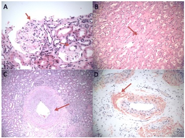
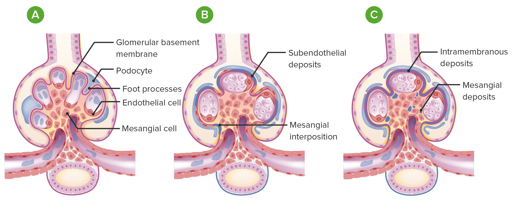
00:01 Moving to the glomerular region. Up until this point, what we have done over and over again is giving you an overview as far as laying down the foundation and then we build upon well. With glomerular diseases, first and foremost, think about where you are, you are in the glomerulus and there are numerous issues or factors that may then result in glomerular disease. Let us begin. First and most importantly at this point, is the fact that your operative word here is biopsy. So the fact that you are taking a biopsy specimen based on the symptoms of your patient. Remember once again. 00:38 Do you remember when we did our session with urinalysis? And with urinalysis, you used information that you had derived based on the history that you got from the patient, either through a clinical vignette or from the attending or what not, and you put all this together so that you can arrive at the proper diagnosis. Well, with the biopsy, there are a couple of important imaging studies that you will have to conduct. You can do an H &E stain and with this, we mean hematoxylin and eosin. And with this, this then allows us to classify the type of glomerular disease. This, for the most part, will be light microscopy. What does light microscopy mean to you? Well, as we go to the various diseases, when it is relevant and pertinent, we will then take a look at diseases in which it is best to classify them through light microscopy. Well, let us say that you don't find exactly as to which you need to or perhaps there is further investigation that is warranted and then you are going to other types of stains and this includes something like immunofluorescence. This is stain number 2, upon what? Biopsy. What was the first one? Light microscopy and we are using H&E stain. 01:48 With immunofluorescence, it is the fact that as the name implies, you are literally immune, well you are looking for the immuno particles and you are fluorescing them and with this particular stain, you probably have seen the color, that bright green type of appearance whenever there is immunoglobulins that are being deposited in your glomerulus. 02:12 Identify proteins and such. 02:14 The linear pattern is something that it is part of your immunofluorescence and let us takes for example you have Goodpasture. From immunology, you have heard Goodpasture being type II hypersensitivity. 02:26 What does that mean to you? It means that it is an antibody dependent issue, isn't it? Well when you are antibody dependent, what does that mean? It means that you are literally attacking your target. So for example, another name for Goodpasture would be anti-glomerular basement membrane antibodies. There are no complexes here that you are forming. Is that clear? No complexes. So what are they doing? These immunoglobulins are then literally attacking the glomerular basement membrane and so therefore if you have these immunoglobulins attacking the basement membrane, therefore, what kind of pattern would you expect to see on immunofluorescence? You are going to light up the glomerular basement membrane in a linear pattern and as we walk through this and I give you specificlly a Goodpasture or antiglomerular basement membrane disease, you will see this pattern under immunofluorescence. Now, if that is attacking the glomerular basement membrane, what if you actually had immune complexes? For example, we have type III hypersensitivity that may occur with SLE. We have type III type hypersensitivity in which what does that mean even? Remember immunology once again. 03:36 It means antigen-antibody complexes. You see the difference. In Goodpasture discussion above, what was it? A type II hypersensitivity. What does that mean? You are attacking the glomerular basement membrane and type III you have immunofluorescence here that is then recognizing the immune complexes of antigen-antibody that is depositing where? Either under the side of the epithelium. When do you say epithelium you tell me what side of the glomerular basement membrane are you on, if I say epithelium? Good, urine side. Whereas if I say subendothelial between the basement membrane and the endothelium, that is the side of the blood. Is that clear? Now, when you have such complexes that can be deposited either underneath the endothelial cell or maybe perhaps under the epithelial cell what have you formed? Immune complexes. What kind of hypersensitivity? Type III. Cannot be detected by electron microscopy and if it cannot so, therefore, what do you do? You do an immunoflorescence in which you find a pattern known as granular. 04:45 So the granular would then be a "lumpy-bumpy." All this means is immune complex deposition. 04:54 If by chance you cannot find certain things under electron microscopy such as your linear pattern, then you'll have to use your immunofluorescence to find that linear pattern. But let us say that you have electron microscopy for the granular pattern, it recognizes immune complexes perhaps and you want further confirmation, then upon immunofluorescence, it is then called your "lumpy-bumpy". Three major patterns of imaging that are important based on the type of pathology that you will get and as we go through both nephritic and nephrotic, I will be asking you this question such as does this weren't require LM, light micrioscopy on H&E? Number 2, biopsy. Immunofluorescence, based on protein deposition and is it a linear pattern or granular pattern? And then the third and final one would be electron microscopy and we had a previous discussion where we went into great detail about electron microscopy where we then identify the glomerular basement membrane, the epithelial side and the endothelial side. 05:58 Let us now continue.
About the Lecture
The lecture Glomerular Disorders by Carlo Raj, MD is from the course Glomerulonephritis.
Included Quiz Questions
Which of the following statements is NOT TRUE regarding Goodpasture’s syndrome?
- It is a type IV hypersensitivity.
- It is a type II hypersensitivity.
- It is an anti-glomerular basement membrane disease.
- It shows a linear pattern on immunofluorescence.
- Antibodies line up evenly against antigens in the glomerular basement membrane.
Which of the following tests would identify patterns and type of protein deposition in glomerular disorders?
- Immunofluorescence stain
- Electron microscopy
- Urinalysis
- Renal ultrasound
- Light microscopy
Which of the following is not true regarding immunocomplex (IC) deposition in the glomerulus?
- Seen as a linear pattern on immunofluorescence stain.
- Seen as a granular pattern on immunofluorescence stain.
- Seen as a "lumpy-bumpy" pattern on immunofluorescence stain.
- It involves antigen-antibody complexes.
- Immunofluorescence stain identifies the pattern of protein deposition.
Author of lecture Glomerular Disorders

Carlo Raj, MD
Customer reviews
5,0 of 5 stars
| 5 Stars |
|
1 |
| 4 Stars |
|
0 |
| 3 Stars |
|
0 |
| 2 Stars |
|
0 |
| 1 Star |
|
0 |
Enthusiastic, to the point, interesting lessons which are enjoyable and educational
