Playlist
Show Playlist
Hide Playlist
Bone Growth

-
Slides 09 Types of Tissues Meyer.pdf
-
Download Lecture Overview
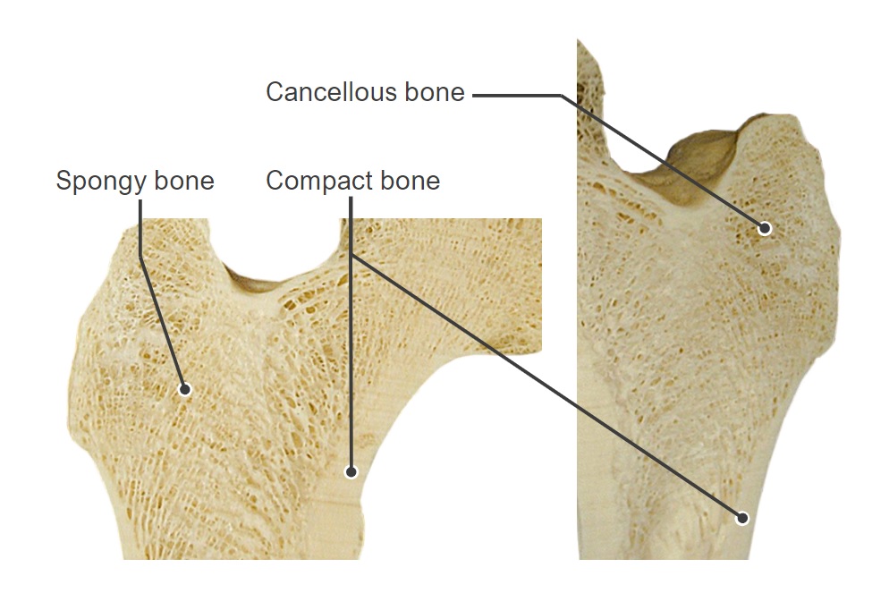
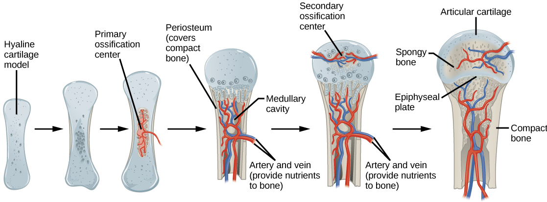
00:00 So how does the bone grow? Having in a fetus have this primary ossification center, initiating bone growth or bone formation, how does that template that cartilage then grow in length and get to the length of the bone that we see in the mature adult? What happens, just at the beginning of birth or at birth, you get the invasion or the same process occurring up at the epiphysis end of the bone or of the developing bone. 00:40 Here you see a section. You see the epiphysial cartilage that represents that cartilage component in the previous sections I have showed you whereby there was this hypertrophied zone of cartilage cells and there was absorption away from that down the diaphysis of the bone creating that medullary cavity. By the time, birth has occurred and all the way through now until adulthood, two things happen. You get a secondary ossification center. You can see that to the right of the epiphysial cartilage. That secondary ossification center develops exactly the same way, suddenly the chondrocytes hypertrophy. They die. They become calcified. The matrix is calcified and bone is laid down and then resolved to some significant degree. This area, this secondary ossification center will develop mostly into spongy bone. Remember that spongy bone we saw on the previous section through the femur I showed you towards the start of this lecture. 01:56 One difference of the secondary ossification center though is that there is no periosteum on the external sides. It remains cartilage because that part of the cartilage and the more distal part of that cartilage is going to be the articular surface of this particular bone. You can see in the far right of this slide another articular surface of articular cartilage. And then what happens is that the bone is going to elongate and widen, but mainly elongate. And it elongates because that epiphysial cartilage component starts to undergo the growth where cartilage cells go through a similar transition through what they did before and bone is being laid down. So just look at those details a little bit more. 02:59 This summarizes what I described in the previous slide. Down the bottom the question is what reminds is its epithelial cartilage template that is going to be the structure that allows the bone to elongate. And when you look this emphysial plate and you see the processes different zone, on the far right-hand side is cartilage. You can see the cells there are just typical cartilage that results on the reserve cartilage. Then you can see where there's higher concentration of chondrocytes. They have divided. They are proliferating and I call those two zones the reserve zone and the proliferation zone, the running away zone and I will describe why I name that in a moment. Then you've got this zone of hypertrophy of cartilage, the calcification of the matrix and then resorption bone is laid down and then resorbed as I described previously. 04:09 Now that running away zone that I mentioned is because when the cartilage matrix is calcified and bone is deposited on it and then resorbed to create the diaphysis, the cavity, those two zones reserves in the proliferation run away from that calcified zone. They keep dividing and moving away, getting away from that calcified area consequent in the bone grows in length or the developing bone grows in length. Same thing happens on the other surface, the secondary ossification center. Cells are continually dividing to run away from the zone of calcified matrix. So the bone elongates. It also thickens, gets wider. 04:59 But bone being laid down form the periosteal collar and then eat their way inside to create a large medullary cavity. The secondary ossification center that I described earlier is mainly occurring not to lengthen the bone, that is done by the epiphysial plate region of proximal to the medullary cavity. The secondary ossification center is mainly to do with the moulding of the shape of the epiphysial head of the bone that I have described again in the start of this lecture and the spongy bone that I showed you. And this shows you some of the processes, little spicules of spongy bone is formed on the cartilage, calcified spicules and is continually removed or remodeled. The bone is red staining here. 05:58 The cartilage is of very pale stain. You see it here in more detail and it just summarize on the left-hand side some of the processes that I've just described as a review for you. 06:14 Later on, at the end of puberty, suddenly all those reserve cartilage cells will suddenly start to disappear. And there will be fusion, there will be a closure of this epiphysial plate and there's just a small evidence of the little line going through there in the adult that you can sometimes see. It is called the epiphysial closure. No more growth of the bone occurs. That is all done by this process.
About the Lecture
The lecture Bone Growth by Geoffrey Meyer, PhD is from the course Bone Tissue.
Included Quiz Questions
Which of the following is not a zone of the epiphyseal plate?
- Zone of regeneration
- Zone of proliferation
- Zone of hypertrophy
- Zone of calcification
- Zone of resorption
The secondary ossification centers generally appear at which of the following sites?
- Epiphysis
- Diaphysis
- Metaphysis
- Periosteum
- Endosteum
Which of the following is the primary site of lengthening of long bones?
- Epiphyseal plate
- Medullary cavity
- Periosteum
- Endosteum
- Diaphysis
Which of the following shows the correct order of bone growth in the growth plate?
- Reserve zone | Proliferative zone | Zone of maturation | Calcified cartilage | Zone of resorption
- Zone of resorption | Reserve zone | Zone of ossification | Proliferative zone | Calcified matrix | Zone of maturation and hypertrophy
- Reserve zone | Zone of maturation and hypertrophy | Zone of resorption | Zone of ossification | Proliferative zone | Calcified matrix
- Reserve zone | Zone of resorption | Calcified matrix | Proliferative zone | Zone of maturation and hypertrophy | Zone of ossification
- Proliferative zone | Zone of ossification | Reserve zone | Zone of resorption | Calcified matrix | Zone of maturation and hypertrophy
Author of lecture Bone Growth

Geoffrey Meyer, PhD
Customer reviews
5,0 of 5 stars
| 5 Stars |
|
5 |
| 4 Stars |
|
0 |
| 3 Stars |
|
0 |
| 2 Stars |
|
0 |
| 1 Star |
|
0 |
