Playlist
Show Playlist
Hide Playlist
Wall Structure of the Small Intestine

-
Slides Wall Structure of the Small Intestine.pdf
-
Download Lecture Overview
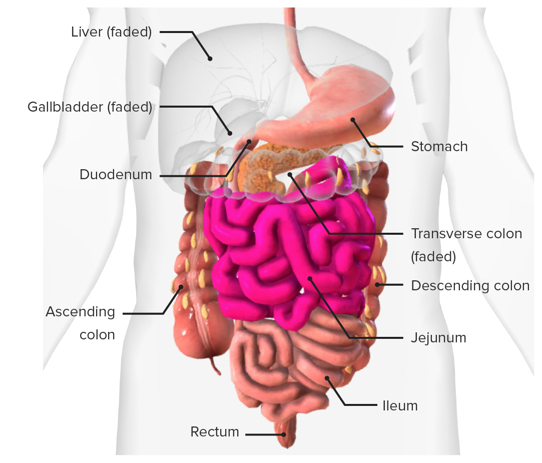
00:01 So let's just quickly have a look at the wall of the small intestine, the microstructure of the small intestine. And we can see again similar to the stomach how various undulations and elevations within the small intestines and these are called plicae. And here we can see these transverse folds running on the internal structure of the small intestine. If we were to have a look at those, you'd see that these undulations are very much increasing the surface area of the small intestine. And we call these intestinal villi. They help to move food along, but they also massively increase the surface area. And actually on the epithelial layer that is on the surface of the intestinal walls on the surface of these plicae, we have microvilli. So on the surface of those epithelial cells, we can see here lining the wall of the small intestine we have microvilli. And they again help the movement of food across this surface and to aid digestion. So let's have a look at the layers of the intestinal wall. We've got those villi that are going out with the microvilli on the epithelial walls. And you can see actually as part of this mucosa in red and blue and in green we have the vasculature that's going to supply those. So each one of those has an arterial supply and a venous drainage, but importantly it also has what's known as a lacteal which is carrying lymph. So here we have the epithelial layer. Deep to the epithelial layer containing these structures we have the lamina propia. We then have an initial mascularis mucosal layer. We have the submucosa. We have a lot of these parenting blood vessels are running along as well before they give off the branches that go towards the villi. And then we have the various longitudinal and circular smooth muscle controlled by the ultimate nervous system that controls and regulates the movement of these walls. Here we can see, like I said, the circular and longitudinal muscle layers. If we were to have a slightly closer look at the villi and the plicae of this region, again you can see the epithelial layer and you can see the blood supply going to and from it. Also here, you can see this lacteal which is a branch of the lymphatic system helping to take lymph to and from this region. Lymph is really important aspect of the digestive tract as it helps to transport additional fluid and white blood cells to and from this region. So having a really detailed lymphatic system is incredibly important. Well, you can also see right to the very bottom of this structure are the intestinal glands or the crypts of Lieberkuhn. And at the bottom here, you have some very specialized cells which really you should have a look at histologicals for more detail about the cells that occupy this space and also the structure and function within the small intestine. But hopefully that's a very high level overview of the microstructure of this area.
About the Lecture
The lecture Wall Structure of the Small Intestine by James Pickering, PhD is from the course Anatomy of the Small Intestine.
Included Quiz Questions
Which part of the gastrointestinal tract is separate from the small intestine?
- Cecum
- Duodenum
- Jejunum
- Ileum
In which segment of the gastrointestinal tract are Brunner’s glands located?
- Duodenum
- Jejunum
- Ileum
- Colon
- Stomach
Which statement regarding the small intestine is inaccurate?
- The plicae disappear with distension.
- The small intestine is 6–7 meters long
- The small intestine has villi and microvilli.
- Ninety percent of nutrient absorption occurs in the small intestine.
- Transverse folds in the small intestine are known as plicae.
Which statement describes the chyme entering the small intestine?
- It is acidic.
- It is basic.
- It is not buffered by pancreatic secretions.
- It has not undergone any digestive processes.
- It passes through the cardiac sphincter.
Author of lecture Wall Structure of the Small Intestine

James Pickering, PhD
Customer reviews
5,0 of 5 stars
| 5 Stars |
|
5 |
| 4 Stars |
|
0 |
| 3 Stars |
|
0 |
| 2 Stars |
|
0 |
| 1 Star |
|
0 |
