Playlist
Show Playlist
Hide Playlist
Vasculature of the Axilla

-
034-03 252309 Axilla and Brachial Plexus-Vasculature-of-the-Axilla.pdf
-
Download Lecture Overview
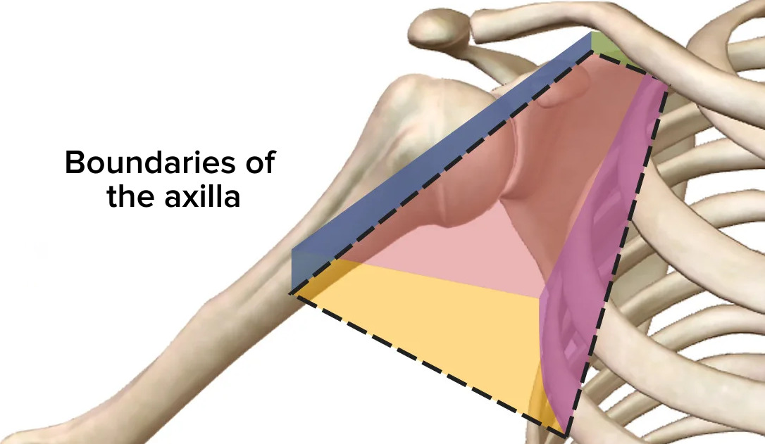
00:01 So, now, let's look at the contents of the axilla. And there's a lot going on within the axilla. 00:07 It's a very busy location. It's full of very important structures connecting the upper limb to the neck and the neck to the upper limb. 00:16 So, first of all, let's have a look at some blood vessels. 00:19 And here, we can see the subclavian artery that's passing from the thoracic cage through the neck into the axilla and it does this by passing along the lateral margin of the first rib. 00:31 And then, enters through the apex of the axilla. 00:34 As it passes through the apex of the axilla, going alongside the clavicle, it becomes the axillary artery. 00:43 It's the same artery, but now, it's running within the axilla, we call it the axillary artery. 00:48 This runs all the way down until the inferior border of the axilla. 00:51 So, we've got the lower border of teres major where it then changes its name again into the brachial artery. 00:58 But notice, this is one continuous blood vessel that changes its name depending on its location. 01:04 So, subclavian, axillary, brachial artery. Here, we can see running anteriorly again, so, forming the anterior wall of the axilla, we have pectoralis minor muscle. 01:15 And that is covering the anterior aspect of the axilla, axillary artery. 01:20 But it also separates the axillary artery into these three parts. 01:25 We've got the first part which is wedged between the clavicle and the superior boundary of pectoralis minor. 01:31 We then, have the second part of the axillary artery which is directly deep to the muscle mass of pectoralis minor muscle. 01:39 And then, the bit that's running from pectoralis minor to the inferior boundary of teres major, we have the third part of the axillary artery. 01:48 And coming off these various parts are various different blood vessels. 01:51 So, we've got three parts of the axillary artery there. 01:56 Coming from the first parts, we have the superior thoracic artery. 01:59 This part is going down to supply some structures on the anterior chest wall. 02:04 And then, coming away from the second part of the axillary artery, we have the thoraco-acromial artery. 02:11 We also have the lateral thoracic artery that starts to run down the lateral aspects of the chest wall. 02:18 If we then, look at the third part of the axillary artery, we find we have the subscapular artery running posteriorly to supply the subscapularis muscle. 02:27 And we also have the anterior circumflex humeral artery running around the surgical neck of the humerus where it's met anastomosis with the posterior circumflex humeral artery. 02:41 So, a high number of blood vessels coming off the axillary artery within those three individual parts. 02:48 We can also trace into the axillary vein, venous blood that's returning from the entire upper limb, all the way eventually to the right side of the heart where we have various blood vessels draining the upper limb. 03:02 So, we've got the brachial veins which are coming deep from the substance of the arm and also, the basilic vein which is draining subcutaneous tissue from the superficial aspect. 03:12 And these converge together and unite to form the axillary vein as it runs in the axilla. 03:18 Also running into the axillary vein but now, much higher up is the cephalic vein. 03:23 And if we remember from the venous drainage of the arm lecture, these are the two principle veins, the cephalic and the basilic that are running on the superficial aspect of the upper limb, draining that subcutaneous tissue. 03:37 The axillary vein then, accompanies the subclavian artery as the subclavian vein running all the way back into the chest cavity as we can see here. 03:46 So, the subclavian artery and the subclavian vein are very closely together, as are the axillary pairing as well. 03:53 And we can see the axillary vein formed from the basilic, the cephalic, and the brachial veins in this diagram here before giving rise to the subclavian vein that returns venous blood to the heart. 04:06 So, closely associated with these venous structures are the lymphatics. 04:10 There's a number of nodes, collections of lymph nodes within the axilla region. 04:16 Here, we have the subscapular node. We have the pectoral nodes. 04:20 We have the central nodes. We have the apical nodes. And we have the infraclavicular nodes. 04:28 These are clusters of lymph nodes located around the axilla and they help to receive and drain the lymphatic fluid from the upper limb and part of the lateral chest region, importantly, the breasts as well. 04:41 These all drain into what's known as subclavian lymph trunk and we have one of these both on the left and the right side. 04:48 You may remember from thoracic lectures that on the left-hand side, lymph is drained by the thoracic duct as well, and here, the left subclavian lymph trunk will drain into the thoracic duct before returning to the venous system. 05:03 It's very important these axillary lymph nodes as they're part of early metastasis of breast cancer. 05:09 So, they help to indicate if breast cancer located within the breast has actually spread to neighboring tissues.
About the Lecture
The lecture Vasculature of the Axilla by James Pickering, PhD is from the course Anatomy of the Axilla.
Included Quiz Questions
Which muscle is associated with the division of the axillary artery into 3 parts?
- Pectoralis minor
- Pectoralis major
- Biceps brachii
- Triceps brachii
- Serratus anterior
The second part of the axillary artery gives off which of the following?
- Lateral thoracic artery
- Superior thoracic artery
- Subscapular artery
- Anterior circumflex humeral artery
- Posterior circumflex humeral artery
Lymph from the posterior thoracic wall and scapula drain into which of the following?
- Subscapular lymph nodes
- Pectoral lymph nodes
- Humeral lymph nodes
- Central lymph nodes
- Apical lymph nodes
Author of lecture Vasculature of the Axilla

James Pickering, PhD
Customer reviews
5,0 of 5 stars
| 5 Stars |
|
5 |
| 4 Stars |
|
0 |
| 3 Stars |
|
0 |
| 2 Stars |
|
0 |
| 1 Star |
|
0 |
