Playlist
Show Playlist
Hide Playlist
Tuberous Sclerosis: Clinical Presentation
-
Strowd CNS Tumors Inherited Tumor Syndromes.pdf
-
Download Lecture Overview
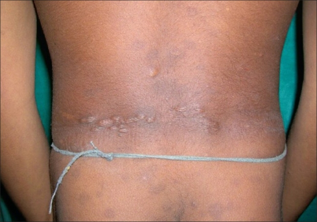
00:01 Here's some examples of some of those findings. 00:03 Again, I really want you to see the ash-leaf macules. 00:06 I want you to know what that hypopigmented spot looks like. 00:09 and that's what's represented in this image, in A. 00:13 Here we see intraoral fibromas, and some dental enamel pits, pits in the teeth, which we can see a young age and patients with tuberous sclerosis. 00:22 And I want you to know what the facial angiofibromas look like. 00:25 And we've seen that a couple of times. 00:26 And here you can see that here hamartomas, in the malar area of the cheek, the nose, the forehead, and around the maxillary portions of the face. 00:36 What other imaging findings do we see in these TSC patients? Well, we know to look at the kidneys, because that's where one of the major tumor types occurs. 00:44 And here in the top left image, you can see multiple tumors in the left kidney and the right kidney. 00:50 These are benign tumors, they're not a cancer, but they can be innumerable and result in loss of kidney function. 00:56 And in those situations should be treated and managed by a multidisciplinary team. 01:01 Here's another appearance of those tumors of the kidneys. 01:03 These are angiomyolipomas or AMLs. 01:09 We also see findings in the lungs. 01:11 This is the one finding in TS that becomes more common as patients age. 01:15 And it's actually more common in women with TSC. 01:18 This is a cystic degeneration of the lungs, that's called LAM. 01:23 And that's the easiest way to remember it. 01:24 And it meets a major criterion for TSC. 01:29 We also see brain lesions. 01:30 And I want you to know that these patients present with seizures, and we should evaluate the brain. 01:35 And there are three findings that you should think about when it comes to brain manifestations of TSC. 01:40 The first is cortical tubers. And that's what you see here. 01:44 They're light lesions, white lesions that are primarily out on the cortex, but can include some of the subcortical fibers. 01:52 They don't enhance with contrast, and are highly epileptogenic. 01:56 The vast majority of patients have multiple cortical tubers spread throughout the brain. 02:00 The anterior portion, the posterior portion, the left and right hemisphere, and many of those contribute to seizures. 02:07 We also see subependymal nodules. 02:09 And this is this the small little white spot right next to the ventricle. 02:12 It doesn't enhance with contrast, it probably doesn't cause a seizure, but is an area of abnormality that needs to be monitored. 02:20 It could grow into a tumor. And if that happens, we would see it enhanced with contrast. 02:25 And the tumor that we see, the third finding that I want you to know about for TSC is a Subependymal giant cell astrocytoma. 02:33 We call these SEGAs. 02:35 And they enhance with contrast, and they very commonly occur in this area that you see here with the green arrow at the foramen of Monro. 02:43 You will recall that this foramen allows the lateral ventricles to drain spinal fluid into the third ventricle. 02:49 It is a critical drainage pipe. 02:51 And if one of those SEGAs obstructs flow through the foramen of Monro patients develop acute hydrocephalus, and can have a big problem quickly. 02:59 So we monitor these closely and if needed treat. 03:04 Here's another example of a Subependymal giant cell astrocytoma. 03:08 In this T1 post contrast image, you can see it lights up with contrast. 03:11 It is an avidly enhancing lesion, and it's sitting right at the area of the foramen of Monro. 03:17 We also see in this patient multiple subcortical tubers, those T2 bright hyperintense lesions spread throughout the brain. 03:24 And we see that SEGA, which is also a T2 bright and enhancing with contrast. 03:31 So how should we monitor these patients? There is surveillance imaging of all organ systems from the top of the head to the bottom of the feet that we need to think about to adequately manage and monitor these patients. 03:42 We watch their dental exam at least every six months to look out for problems with dentition, which is not uncommon. 03:48 We think about neuropsychiatric and neurocognitive function annually with a comprehensive test, and more commonly in patients who have some type of abnormality. 03:57 Neurologic manifestations need to be monitored as well. 04:00 We do EEG or electroencephalography, to evaluate for seizures and MRI of the brain about every one to three years to look for certain brain findings and specifically those concerning tumors.
About the Lecture
The lecture Tuberous Sclerosis: Clinical Presentation by Roy Strowd, MD is from the course CNS Tumors.
Included Quiz Questions
Which of the following is an oral finding of tuberous sclerosis?
- Intraoral fibroma
- Oral aphthous ulcer
- Leukoplakia
- Oral squamous cell carcinoma
- Glossitis
Which of the following is a renal finding of tuberous sclerosis?
- Angiomyolipoma
- Renal cell carcinoma
- Renal papillary necrosis
- Rhabdomyolysis
- Nephrolithiasis
Which of the following is one of the recommended surveillance tests for patients with tuberous sclerosis?
- EEG
- Biopsy
- Ophthalmoscopy
- Electromyography
- Nerve conduction studies
Customer reviews
5,0 of 5 stars
| 5 Stars |
|
5 |
| 4 Stars |
|
0 |
| 3 Stars |
|
0 |
| 2 Stars |
|
0 |
| 1 Star |
|
0 |


