Playlist
Show Playlist
Hide Playlist
Temporal, Infratemporal and Pterygopalatine Fossae

-
Slides Anatomy Temporal Infratemporal Pterygopalatine Fossae.pdf
-
Download Lecture Overview
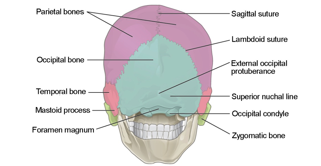
00:01 Now we're going to look at some shallow depressions or Fossae in the skull called the temporal, infratemporal and pterygopalatine fossae. 00:13 We'll start by looking at the temporal fossa. 00:17 Medially, we have the parietal bone, and frontal bone, and sphenoid bone, as well as the squamous portion of that temporal bone. 00:28 Laterally is the fascia called the temporal fascia that's going to attach here. 00:37 Anteriorly, we have the zygomatic process of the frontal bone and the frontal process of the zygomatic bone. 00:49 Superiorly, there is a line called the temporal lines which serve as attachment for muscle and fascia. 00:57 Inferiorly, we have that zygomatic arch and in this fossa, we're mostly going to find a very large muscle of mastication called the temporal muscle or temporalis. 01:13 There are also going to be vessels called the deep temporal arteries and veins. 01:18 The zygomatic temporal nerve and a very important branch of the external carotid, one of the terminal branches. 01:27 The superficial temporal artery. 01:31 We see various branches as well such as the middle temporal artery. 01:37 The infratemporal fossa is going to be just below the temporal fossa. 01:43 And so we're going to take an inferior view from the side. 01:47 We can see the roof of the fossa here being composed of the inferior surface of the greater wing of the sphenoid. 01:57 Here's some more of the temporal bone and we see the communication with the temporal fossa underneath this arch. 02:07 We see the posterior surface of the maxilla more anteriorly, as well as the inferior orbital fissure. 02:16 Medially, we have something called the pterygomaxilliary fissure and the pterygoid process. 02:26 We also see a little bit of the pharynx from here. 02:30 Laterally, we have that vertical process of the mandible that we call the Ramus. 02:36 Let's look at what is in this fossa. 02:41 So we have a ligament that's keeping our jaw connected to the rest of our skull that's sphenomandibular ligament. 02:50 We also have a muscle mastication in this area. 02:52 We see called the lateral pterygoid muscle. 02:56 We also have the other pterygoid muscle that's more medially the medial pterygoid muscle another muscle of mastication. 03:05 We also see in this area, the mandibular nerve or the third branch of trigeminal. 03:12 We have the buccal nerve coming off of it. 03:15 The lingual nerve, the alveolar nerve. 03:20 The auriculotemporal nerve. 03:23 And then this little thing here called the chorda tympani which is actually a portion of cranial nerve seven that's essentially hitching a ride with the mandibular nerve in order to reach its destination. 03:36 So the chorda tympani is actually a way for sneaking some cranial nerve seven fibers along the lingual nerve to reach the tongue. 03:46 In terms of arteries, here we see the external carotid giving rise to the maxillary artery branch around the same time that is giving rise to the superficial temporal artery. 03:58 We see some branches off of the maxillary artery as well including the inferior alveolar artery that travels along with the inferior alveolar nerve through the mandibular canal. 04:10 We see the middle meningeal artery coming off the maxillary which again will enter the skull in order to supply the meninges. 04:18 We also see the pterygomeningeal artery, as well as the posterior deep temporal artery. 04:27 We also have a pterygoid artery closer to the area of the pterygoid muscles, the buccal artery in the cheek area. 04:37 And we have an anterior deep temporal artery as well. 04:40 So that temporal fossa is very well supplied. 04:45 We also see a descending palatine artery in the area of the hard palate. 04:51 There's a superior alveolar artery doing the same job as the inferior alveolar artery providing blood apply to the upper teeth. 05:01 In terms of venous drainage, we have a very large plexus of veins called the pterygoid plexus. 05:09 The next fossa is the pterygopalatine fossa, which gives you some idea of where it's going to be located, somewhere in those areas of the pterygoid muscles and plates and the palate. 05:21 So we zoom in, we can see the anterior wall here, which is the posterior aspect of the maxilla. 05:30 And we can see a little bit of the sphenoid bone hanging down here as well. 05:36 We see a little bit of the palatine bone, which is actually a small portion of the hard palate. 05:47 There are a few ways to get to the pterygopalatine fossa. 05:51 Here we see the inferior orbital fissure that we saw from an anterior view of the skull a little while back. 05:59 We have the pterygomaxillary fissure in that space between the pterygoid and the maxilla. 06:06 We have the sphenopalatine foramen, which also gives you an idea of where we're located. 06:13 There is a little narrow opening here in the area of the palate called the palatine canal. 06:19 And then there's also an opening of the skull, we already saw called the foramen rotundum. 06:26 Similar in the area of the pterygoid plates, we have the pterygoid canal and a palatovaginal canal.
About the Lecture
The lecture Temporal, Infratemporal and Pterygopalatine Fossae by Darren Salmi, MD, MS is from the course Skull.
Included Quiz Questions
Which muscle lies in the temporal fossa?
- Temporalis
- Zygomaticus
- Frontalis
- Submandibular gland
- Parotid gland
What is part of the roof of the infratemporal fossa?
- Inferior surface of the greater wing of the sphenoid
- Posterior surface of the maxilla
- Inferior orbital fissure
- Pterygoid process
- Pharynx
What forms the lateral boundary of the infratemporal fossa?
- Ramus
- Posterior surface of the maxilla
- Temporal bone
- Greater wing of the sphenoid
- Lesser wing of the sphenoid
What are the gateways to the pterygopalatine fossa? Select all that apply.
- Inferior orbital fissure
- Sphenopalatine foramen
- Pterygomaxillary fissure
- Palatine canal
- Foramen magnum
Author of lecture Temporal, Infratemporal and Pterygopalatine Fossae

Darren Salmi, MD, MS
Customer reviews
5,0 of 5 stars
| 5 Stars |
|
5 |
| 4 Stars |
|
0 |
| 3 Stars |
|
0 |
| 2 Stars |
|
0 |
| 1 Star |
|
0 |
