Playlist
Show Playlist
Hide Playlist
Surface Features of the Liver

-
Slides Surface Features of the Liver.pdf
-
Download Lecture Overview
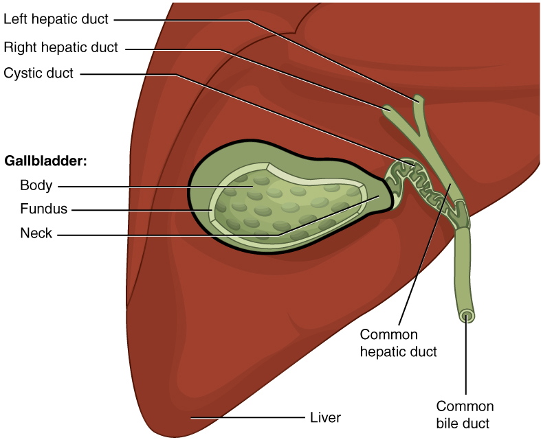
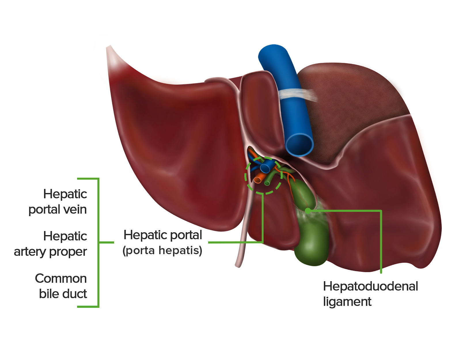
00:01 So let's have a look at the liver itself in isolation. 00:03 If we were to take it out of the abdomen, we can see we have a very large anterior surface that really runs alongside the anterior lateral abdominal wall and also part of the diaphragm. 00:13 We have the right surface and we have a superior surface very much nestled along the underside of the diaphragm. 00:20 If we were to spin the liver around, we'd see its posterior surface. 00:24 And then the inferior surface. 00:26 Delivers a strange shape where it has kind of a sloping diagonally referenced inferior surface, as opposed to a more flattened posterior surface that sits quite tightly against the diaphragm. 00:38 So we have both aspects of the liver, which we can see there anterior surface and posterior surface, a slanting diagonally orientated inferior surface. 00:47 And there's the superior and right surfaces as well. 00:51 This large anterior surface that I've alluded to really does touch on all of the diaphragm. 00:55 So we have the diaphragmatic surface. 00:57 And all of that posterior and inferior surface is intimately associated with such things as the kidney, the transverse colon, the hepatic flexure, which we spoke about, and things like the stomach, and it's known as the visceral surface. 01:11 So, these are in important boundaries to remember now we can see in the green line, the inferior border. 01:18 If you were to have a look at this anterior surface of the liver, we see that it can be split into two lobes, the right lobe and the left lobe, as you can see there. 01:26 And they are partitioned by this falciform ligament. 01:30 Again, a remnant of the ventral mesentery during embryo logical development. 01:34 And a very important connection there is the round ligament that runs down the anterior abdominal wall towards the umbilicus. 01:41 And again, that was an important entry point for blood to pass into the developing embryo and fetus during development. 01:48 That's the round ligament on the free edge of the falciform ligament there. 01:53 So we can see the anterior surface of the liver there, if we were to rotate the liver upwards, so we take that inferior border and flip it to the top of the screen. 02:03 We then left with this view. 02:05 So it's kind of looking at upside down. 02:07 But it's as if you're looking at the cadaver, looking at the liver and you've got your fingers under this inferior border and your reflected up. 02:14 As you take that up, you'll see the gallbladder coming up with it. 02:17 We can see that sitting next to what we call the quadrant lobe. 02:21 And then we can see this small fissure that's running between the quadrant lobe and the left lobe. 02:27 And that's the fissure for the ligamentum teres. 02:29 And that again, is an important and biological remnant. 02:32 We've got the gallbladder sitting there, and we've got the porta hepatis, which you may remember has various structures passing through it. 02:39 So it's connected to the free edge of the lesser omentum. 02:42 So, the bile duct, the hepatic portal vein, and the hepatic artery. 02:47 A small lobe that's just situated in between the right lobe and the left lobe, we have the caudate lobe here, and that is sitting next to the inferior vena cava, that large structure in blue. 02:58 Between the caudate lobe and the left lobe really running towards this fissure of ligamentum teres. 03:04 We have the fissure of ligamentum venosum. 03:07 And these, again are very important and biological structures that are part of that developmental process. 03:12 So you should go and look up what those terms mean is part of the blood flowing from the umbilicus through the liver and then into the general circulation of the developing embryo and fetus. 03:23 If we just stay looking at this surface for a moment, or two, the porta hepatis is important. I mentioned it previously. 03:28 But it is worth going over it again. 03:30 Porta Hepatis is passing into the liver. 03:33 So, Porta Hepatis is this opening that goes into the liver and it's full of that portal triad. 03:39 Those three structures. 03:40 The portal hepatic vein, taking kind of poorly oxygenated but nutrient rich blood from the gastrointestinal tract to the liver for it to be processed and then joined general circulation by hepatic veins. 03:55 It's important to remember that it is not an hepatic vein. 03:57 This is a portal hepatic vein taking poorly oxygenated but nutrient rich blood from the gastrointestinal tract into the liver. 04:06 We also have a hepatic arteries which are coming from the hepatic artery that comes from the celiac trunk when we had the hepatic artery proper, or the common hepatic artery that passed towards the liver, and then it's passing into the various areas of the liver, the various different lobes got a hepatic arteries and important we've got the hepatic bile ducts, which is taking bile out of the liver, and passing into the biliary tree, which we'll see in a moment or twos time. 04:32 So it's important we have an understanding of this area of the liver. 04:36 Important as well that remember, there's a lot of organs that have the liver running up against them. 04:42 So we can see here we have the duodenum, the large intestine, the kidney, the esophagus, the stomach, all of these are imparting various different projections onto the liver, indicating the position in which the liver sits within the abdomen. 04:58 So you can see how the posterior and inferior surface of the liver this residual surface has all of the structures closely associated with it the duodenal, the colic impression, which is around the hepatic flexure the renal impression for the kidney, esophageal impression, which is the esophagus and the gastric impression which is around the stomach. 05:17 Important to appreciate these organs are touching on this inferior visceral surface of the liver.
About the Lecture
The lecture Surface Features of the Liver by James Pickering, PhD is from the course Anatomy of the Liver and Gallbladder.
Included Quiz Questions
Which blood vessel has a remnant that is found in the free edge of the falciform ligament?
- Umbilical vein
- Umbilical artery
- Hepatic vein
- Hepatic portal vein
Which lobes of the liver are closely related to the gallbladder?
- Right and quadrate
- Left and quadrate
- Caudate and quadrate
- Left and right
Which lobes of the liver is the porta hepatis located between?
- Caudate and quadrate lobes
- Left and right lobes
- Left lobe and caudate lobe
- Right lobe and caudate lobe
- Left lobe and quadrate lobe
In which ligament is the portal triad present?
- Hepatoduodenal ligament
- Falciform ligament
- Round ligament
- Coronary ligament
- Hepatogastric ligament
Author of lecture Surface Features of the Liver

James Pickering, PhD
Customer reviews
5,0 of 5 stars
| 5 Stars |
|
5 |
| 4 Stars |
|
0 |
| 3 Stars |
|
0 |
| 2 Stars |
|
0 |
| 1 Star |
|
0 |
