Playlist
Show Playlist
Hide Playlist
Superior Mesenteric Artery

-
Slides Superior Mesenteric Artery.pdf
-
Download Lecture Overview
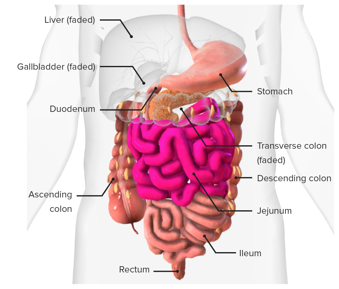
00:01 So now let's talk about the superior mesenteric artery. 00:05 we have the abdominal aorta, which is passed through the diaphragm giving rise to the superior mesenteric artery. 00:12 And the superior mesenteric artery appears at a roundabout the first lumbar vertebrae. 00:18 Just above where the superior mesenteric artery leaves the aorta, we actually have the splenic vein, and that is draining blood across the aorta from the spleen ultimately going to form the hepatic portal vein. 00:31 We see here the complex anatomy of this region in that the hepatic portal vein is formed very much underneath the neck of the pancreas receiving branches from the splenic vein. 00:43 And as we'll see later, it receives branches from the superior mesenteric vein and also the inferior mesenteric vein. 00:51 But we can add some more complications here where we can see the inferior vena cava has now been added directly underneath the head of the pancreas. 01:00 And we can see feeding into this area by looping underneath the superior mesenteric artery. 01:05 We have the left renal vein draining the left kidney. 01:09 This is a really complicated area, and we can see how by adding in the uncinate process of the pancreas this region is really complicated and actually trying to do any surgery to the pancreas can be really difficult because of this complex anatomy. 01:23 By adding in the duodenum, you can see how the relationship of the superior mesenteric artery appears from posteriorly, emerges through the space around the uncinate process of the pancreas to then sit anterior to the duodenum. 01:38 This is a really complex area of anatomy, and now we're adding in the stomach, you can see it better. 01:43 The organization of the superior mesenteric artery emerging from the aorta. 01:49 As we touched on when we looked at the celiac trunk and we added these in for completion, we have that transition point between the foregut and the midgut. 01:58 So here we can see coming off the superior mesenteric artery, we can see the anterior pairs of the superior and inferior pancreaticoduodenal arteries. 02:08 So we can see coming off the superior mesenteric artery is the inferior pancreaticoduodenal arteries, anterior version, and that's going to anastomose with the anterior version from the superior pancreaticoduodenal artery. 02:23 We can also see the inferior pancreaticoduodenal arteries posterior version. 02:29 And as we should be familiar now, that is going to customers with the posterior version of the superior pancreaticoduodenal artery. 02:38 And this is coming from the celiac trunk by way of the gastroduodenal artery. 02:43 So we can see that complex anastomosis. 02:46 Once again, around the head, uncinate process, and duodenum the transition between the foregut and the midgut. 02:54 If we then continue with the superior mesenteric artery very much moving away to the left running within the mesentery of the small intestine, we have the jejunal arteries, and we have the ileal arteries. 03:06 And as they pass towards the substance of the small intestine, so they pass towards the jejunum and ileum. 03:13 They form these anastomosing arches or these arterial arcades. 03:18 And they end up increasing the length of this arterial network by essentially running on the inside of the small intestines but running parallel to it. 03:28 So then we have a paralleling blood vessel run alongside the small intestine. 03:35 Coming off this parallel blood vessel, we have a series of straight arteries, and these run directly into the small intestine, the jejunum and ilium itself, and we call these vasa recta. 03:48 So coming off the jejunal, and the jejunal arteries, and the ileal arteries, we have a series of these anastomosing archers called arterial arcades. 03:57 Coming from them a series of straight arteries called vasa recta. 04:02 If we then take away the small intestine and start adding in the large intestine, we can start to see some very important structures. 04:10 Again, we have the superior mesenteric artery, and then we have a series of blood vessels coming off the superior mesenteric artery. 04:17 Here we have the middle colic, and it has a couple of branches destined towards the transverse, the hepatic flexion region of the transverse and the ascending colon. 04:29 We have the right colic artery. 04:31 Again, we have an ascending and a descending branch going up the way and down around the ascending colon. 04:37 We also have the ileocolic artery. 04:40 Here we have the colic branch going to the colon and the ileal branch going to supply the ileocecal junction. 04:46 We also have an appendicular branch that is going to supply the appendix. 04:51 This is a classic textbook version of what it looks like. 04:54 It may be very different if you have access to looking at a cadaver. 04:57 Or if you will have to having access to a patient. 05:00 Their blood supply may not be quite what it looks like in the textbook, but it's good to have this understanding to start with. 05:08 As mentioned previously, when we looked at the arterial arcades running around the small intestine, we have a very similar approach in the large intestine. 05:16 But here we call it the marginal artery. 05:19 So we have contributions from the middle, right, ileocolic that are running towards the large intestine. 05:25 And then just as they get there, they form this connecting loop that runs on the inside of all of the large intestine, and that's known as the marginal artery. 05:35 Coming off it we have those straight arteries. 05:37 Again, to supply the tissue itself, and these are the vasa recta. 05:41 So let's go back to our diagram. 05:43 Starting with the superior mesenteric artery, we have the pancreaticoduodenal artery. 05:49 And they give rise to anterior and posterior branches. 05:53 These are going to anastomose with anterior and posterior versions of the superior pancreaticoduodenal artery. 06:01 And these are coming from the celiac trunk. 06:03 So here we have the anastomosis between the foregut and the midgut. 06:09 We then look at the superior mesenteric artery, which gives rise to jejunal and ileal arteries. 06:15 Then coming away to the right we have the middle colic, left colic, right colic, ileocolic arteries. 06:23 We can see there, the left colic artery and the middle colic artery are really important as they're going to form another anastomosis but this time around the midgut and the hindgut, and we can see the left colic artery is going to eventually come from the inferior mesenteric artery. 06:39 And we'll see that in a moment or two. 06:41 Just for completion, coming off the ileocolic artery you can see at the bottom is the appendicular artery that goes on to supply the appendix. 06:50 Again, maybe try and recreate this in a simple drawing and see if you get it right and keep practicing and to get all the arteries connected in the right way.
About the Lecture
The lecture Superior Mesenteric Artery by James Pickering, PhD is from the course Vascular Supply of the Abdomen.
Included Quiz Questions
A person has an atherosclerotic plaque in the superior mesenteric artery that has completely occluded the blood vessel. Which part of the gastrointestinal tract will show no effects from this occlusion?
- Distal 1/3 of the transverse colon
- Jejunum
- Ileum
- Appendix
- Ascending colon
Author of lecture Superior Mesenteric Artery

James Pickering, PhD
Customer reviews
5,0 of 5 stars
| 5 Stars |
|
5 |
| 4 Stars |
|
0 |
| 3 Stars |
|
0 |
| 2 Stars |
|
0 |
| 1 Star |
|
0 |
