Playlist
Show Playlist
Hide Playlist
Superficial Layer of the Posterior Compartment of the Leg
-
Slide Superficial Layer of the Posterior Compartment.pdf
-
Download Lecture Overview
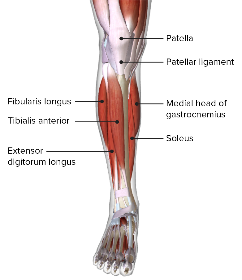
00:01 Now let's move to the posterior compartment of the leg and start by looking at the superficial layer. 00:09 So in the posterior compartment of the leg, we have a superficial and a deep layer. 00:13 Let's start by looking at the superficial layer where we find gastrocnemius and then deep to gastrocnemius, we find soleus. 00:21 And then we'll also find plantaris as well. 00:24 Before we move on to plantaris, soleus and gastrocnemius are two important muscles because they form the most substantial parts of the calcaneal tendon and we can sometimes call these the triceps surae. 00:36 Here we can see the calcaneal tendon in green is been formed by both the soleus and the gastrocnemius muscles as it passes most superficially towards the calcaneal bone. 00:47 Here we can see plantaris by plantar which doesn't really blend with the calcaneal tendon, although it does run alongside its medial aspect, which we can see there. 00:58 So now let's start by looking at the most superficial of these muscles, which is gastrocnemius. 01:03 Gastrocnemius has two heads and its first head is coming from the posterior aspect of the medial condyle of the femur. 01:11 This is the medial head, and then the lateral head comes from a very similar position but on this lateral aspect. 01:17 It comes from the posterior aspect of the lateral condyle of the femur. 01:21 So we have a medial and lateral head. 01:24 If we then look at soleus, we see soleus coming from the head of the fibular, and also that soleal line which we saw on the tibia. 01:33 Both of these muscles converge down onto that calcaneal tendon, which goes to the posterior surface of calcaneus. 01:40 That large substantial posterior bone within the foot. 01:46 If we then look at the calcaneal tendon, we can see those fibers right in the medial part here, these are formed mostly by soleus. 01:54 Whereas the more laterally positioned fibers of calcaneal tendon is formed by gastrocnemius. 02:00 So now let's turn our attention to plantaris. 02:03 Plantaris which is a small muscle with a very long tendon and its muscle belly is located in the popliteal fossa, behind the knee. 02:11 We can see plantaris here, it's originating from the lateral supracondylar line, and then it passes all the way down towards the calcaneus where it merges somewhat with the calcaneal tendon. 02:22 It doesn't form as tight and adhesion to the calcaneal tendon as gastrocnemius and soleus does but it runs down this medial aspect of it. 02:32 Innervation of this posterior compartment superficial layer is why the tibial nerve and remember the tibial nerve is formed from the bifurcation of the sciatic into the tibial and the common fibular nerves. 02:45 The function of the posterior compartment muscles superficial layer, so soleus, gastrocnemius and plantaris is very much one of plantar flexion of the foot. 02:56 So helping you to stand on your tiptoes. 02:59 As it also passes across the knee joints, it can help to flex the knee, but this is only gastrocnemius and plantaris, because they crossed the posterior aspect of the knee joint. 03:09 Soleus, which doesn't cross the knee joint doesn't take part in this function. 03:13 But these muscles gastrocnemius and plantaris can help flexion of the knee.
About the Lecture
The lecture Superficial Layer of the Posterior Compartment of the Leg by James Pickering, PhD is from the course Anatomy of the Leg.
Included Quiz Questions
What are the muscles of the superficial posterior compartment of the leg? Select all that apply.
- Soleus
- Gastrocnemius
- Fibularis longus
- Fibularis brevis
- Fibularis tertius
What is the origin of the soleus?
- Head of fibula
- Posterior aspect of lateral condyle of femur
- Posterior aspect of medial condyle of femur
- Base of 1st metatarsal
- Base of 5th metatarsal
Customer reviews
5,0 of 5 stars
| 5 Stars |
|
5 |
| 4 Stars |
|
0 |
| 3 Stars |
|
0 |
| 2 Stars |
|
0 |
| 1 Star |
|
0 |


