Playlist
Show Playlist
Hide Playlist
Superficial Layer of the Anterior Compartment of the Forearm

-
Slide Superficial Layer of AC of Forearm.pdf
-
Download Lecture Overview
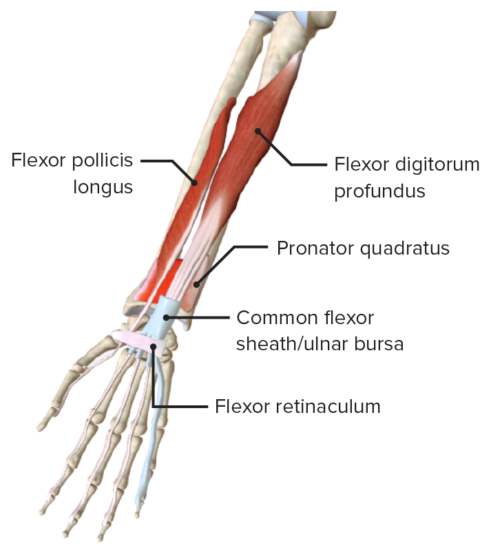
00:01 So, now, let's move on to a specific compartment in the forearm. 00:04 And this is the anterior compartment. So, let's have a look at the anterior compartment. 00:09 And really, we can see there's a whole series of muscles. 00:12 And they're divided into a number of layers in this anterior compartment. 00:17 So, here, we're looking at a right forearm, looking at its anterior surface and we're looking at the most superficial layer. 00:24 You could see there's a number of muscles which are really running away from the medial epicondyle. 00:30 Pronator teres, flexor carpi radialis, palmaris longus, flexor carpi ulnaris. 00:38 We can see four muscles running away in this superficial layer on the anterior compartment of the forearm. 00:45 The middle layer has one large substantial muscle, flexor digitorum superficialis. 00:53 And that sits in the middle layer of these forearm muscles. 00:57 We then, have a deep layer, so, superficial, middle, now, a deep layer, and this deep layer is flexor digitorum profundus and flexor pollicis longus. 01:07 We can also see deepest of them is pronator quadratus. 01:12 So, we can see a whole series of these muscles running within the deep aspect of the forearm. 01:17 If we now return to the superficial layer of muscles within the anterior compartment of the forearm, pronator teres, flexor carpi radialis, palmaris longus, flexor carpi ulnaris, we can start looking at them in more detail. So, here's pronator teres. 01:34 Pronator teres has a humeral head and it also has an ulnar head. 01:39 Humeral head's coming from the humerus, ulnar head coming from the ulna. 01:43 They pass all the way down from the medial epicondyle or the coronoid process, depending on it being the humeral head or the ulnar head. 01:52 So, the humeral head is coming from the medial epicondyle. 01:55 The ulnar head is coming from the coronoid process on that bone. 02:00 And they pass all the way to the lateral surface. That's important. 02:03 The lateral surface of the radius, about halfway down along the shaft of the radius. 02:10 And this is important in pronating. That's why it's important in it attaching to the lateral surface of the radius, so, it helps to flex the arm, yes, flex the forearm at the elbow joint but importantly, it helps with pronation of the forearm. 02:26 We've heard previously about supinator muscle and supination. 02:30 This does the opposite. So, from a supinated position, it helps to pronate the forearm, hence, its name pronator teres. 02:39 It also has an insignificant function but an important one, I suppose, on flexing the forearm at the elbow joint. 02:46 Now, let's move on to flexor carpi radialis. Flexor carpi radialis is an important muscle that sits on the radial aspect of the forearm as we can see. 02:56 Again, it comes from the medial epicondyle and it passes all the way to the base of the second metacarpal within the hand. 03:05 It's an important muscle and it's associated with flexion of the wrist. 03:10 So, flexor carpi radialis, important in flexing the wrist. 03:13 But it also, because it's running on this lateral aspect, helps in abducting the wrist. 03:21 So, moving the wrist away from the midline. 03:23 So, flexor carpi radialis, flexion of the wrist and abduction of the wrist. 03:29 Palmaris longus is another muscle within the superficial layer. 03:33 It also comes from the medial epicondyle and it runs all the way through the forearm to attach to the flexor retinaculum and the palmar aponeurosis. 03:42 It doesn't really attach to a bone but it blends with some very tough fibrous structures within the hand, the flexor retinaculum and the palmar aponeurosis. 03:52 We'll come back to those structures when we look at the hand in more detail in later section. 03:57 So, the function of palmaris longus is similar to other muscles in the superficial layer in that it helps to flex the wrist. 04:05 But it also helps to create tension in the palmar aponeurosis and that's important in helping to maintain a firm grip when you're holding things like screwdrivers, for example. 04:15 It helps to create tension in the palmar aponeurosis. 04:19 One of the muscles in the superficial layer that we can come to next is flexor carpi ulnaris. 04:24 Flexor carpi ulnaris also comes from the medial epicondyle. We can see it here. 04:29 But it comes a little bit more from the posterior aspect, partly because there's already three muscles on the medial epicondyle and this is then situated more posteriorly but still coming from the medial epicondyle. 04:42 It runs down the medial aspect of the forearm, run alongside the ulna, where it goes and attaches to a number of structures within the wrist and the hand. 04:51 The pisiform, the hook of the hamate, and the fifth metacarpal. 04:55 So, now, let's look at the function of flexor carpi ulnaris which is very similar to flexi carpi radialis except because it's now located on the medial aspect of the forearm, it serves to adduct the wrist. 05:08 So, we can still flex the wrist similar to other muscles in this superficial layer. 05:13 But instead of abducting the wrist, now, this one adducts the wrist due to its location on the medial aspect of the forearm and its insertion into those bony structures in the wrist and the hand, important adductor of the wrist. 05:30 Now, let's look at the innervation of this superficial layer. 05:33 We can see here, we've got the ulnar nerve which is passing posterior to the medial epicondyle of the humerus. 05:40 And here, we've got flexi carpi ulnaris for helping us to locate ourselves. 05:45 Here, we can see the median nerve is running down, giving branches to pronator teres, branches to flexor carpi radialis, and giving a branch to palmaris longus. 05:55 But importantly, highlighted in blue, flexor carpi ulnaris is not supplied by the median nerve, it's supplied by the ulnar nerve which we can see here. 06:04 So, two principal nerves, flexi carpi ulnaris most medially supplied by the ulnar nerve whereas the median nerve is supplying pronator teres, flexor carpi radialis, and palmaris longus.
About the Lecture
The lecture Superficial Layer of the Anterior Compartment of the Forearm by James Pickering, PhD is from the course Anatomy of the Forearm.
Included Quiz Questions
Which muscle inserts onto the base of the second metacarpal bone?
- Flexor carpi radialis
- Flexor carpi ulnaris
- Palmaris longus
- Pronator teres
- Flexor digitorum superficialis
Which muscle is innervated by the ulnar nerve?
- Flexor carpi ulnaris
- Flexor carpi radialis
- Flexor digitorum superficialis
- Pronator teres
- Palmaris longus
Author of lecture Superficial Layer of the Anterior Compartment of the Forearm

James Pickering, PhD
Customer reviews
5,0 of 5 stars
| 5 Stars |
|
5 |
| 4 Stars |
|
0 |
| 3 Stars |
|
0 |
| 2 Stars |
|
0 |
| 1 Star |
|
0 |
