Playlist
Show Playlist
Hide Playlist
Small Intestine: Duodenum

-
Slides 15 Human Organ Systems Meyer.pdf
-
Download Lecture Overview
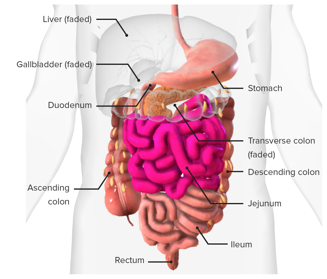
00:00 glial cells, supportive cells. The small intestine has three different components, and they're indicated on this slide. On the left-hand side is the duodenum. The duodenum is characterized by having submucosal glands, mucous secreting glands, particularly near the entrance from the pylorus component of the stomach where acidic chyme could be entering into the duodenum. These mucus-secreting glands try to neutralize any of that acid. And these submucosal glands will become very limited as you progress along the length of the duodenum. 00:45 The jejunum has uncharacteristic features, but really, the only difference between the duodenum and later the ileum is the fact that these have very long villi, but they don't have submucosal glands, and they don't have Peyer's patches as the ileum has. 01:10 Peyer's patches are areas that give an indication that there has been an immune response occurring in response to the invasion and identification of a pathogen. So, using these criteria, you can see or at least identify and distinguish the duodenum from the jejunum and the ileum. 01:33 And they are the three main components as I mentioned before of the small intestine. 01:39 These two sections show examples of the gut-associated lymphatic tissue. There's also a similar lymphatic tissue in bronchi of the lung, the bronchi-associated lymphatic tissue. And sometimes we see this lymphatic tissue underneath other mucosal surfaces. So we call them mucosa-associated lymphatic tissue. But as I said previously, they're evidence of an immune response.
About the Lecture
The lecture Small Intestine: Duodenum by Geoffrey Meyer, PhD is from the course Gastrointestinal Histology.
Included Quiz Questions
Brunner's glands produce which of the following types of secretions?
- Mucus-rich alkaline secretions
- Mucus-rich acidic secretions
- Serous alkaline secretions
- Serous acidic secretions
Author of lecture Small Intestine: Duodenum

Geoffrey Meyer, PhD
Customer reviews
5,0 of 5 stars
| 5 Stars |
|
5 |
| 4 Stars |
|
0 |
| 3 Stars |
|
0 |
| 2 Stars |
|
0 |
| 1 Star |
|
0 |
