Playlist
Show Playlist
Hide Playlist
Scalene Muscles

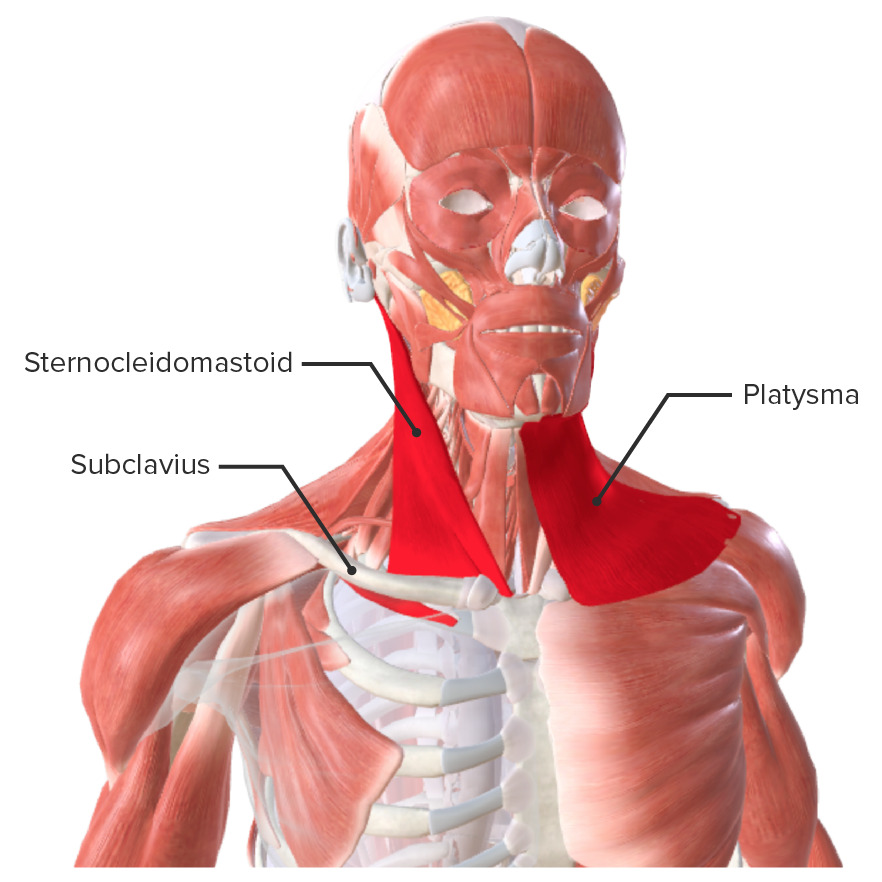
00:00 coming from C7 on its own. And the lower trunk is C8, T1. So if you imagine here at this level, you have the C5, C6, C7, C8, T1. That’s how it comes out, and they are forming the trunk. Upper, middle, lower. The favorite anatomical question is these trunks lie between two muscles, what are they? Scalenus medius and scalenus anterior. 00:30 Scalenus anterior, okay. So you have the scalene muscles like this coming from the neck, from the transverse process of the vertebra. And these nerve roots are coming out between the scalene. So when you do an interscalene block, this is where you’re going. 00:47 What’s the function of scalene muscles? What do they do? Where do they originate? Where do they insert? And why is it important? They insert on the clavicle? Yeah, very good, the scalene tubercle in the clavicle as well, yup, as well as the first trip. How many scalene muscles are there? Three. 01:08 Three. So you have scalenus anterior, medius in posterior. Why are they called scalene? What does scalene mean? What is a scalene triangle? When you have an isosceles triangle, equilateral triangle, what does a scalene triangle mean? Never heard of it before. Never heard of a scalene triangle! No. Unequal sides. So you have two sides, the third side is longer than the other two. So that’s why it’s called scalene. So the three scalene muscles are of different lengths. They’re all starting from the transverse process of the cervical vertebra. You don’t have to know the insertion of the individual ones, but you need to remember that from the neck, it is coming to the clavicle at the top and the first rib, as well as to the second rib. So that’s why it’s one of the accessory muscles of respiration. When you inspire, these ribs are pulled up because of scalene muscles. That’s action of scalene, and this is your anatomy relation, the roots of the plexus come in between the anterior. Scalenus anterior and scalenus medius. 02:21 So we are here now. We are in the upper trunk, middle trunk, lower trunk. These trunks lie in the posterior triangle of the neck, the clavicular region. These are your three trunks lying in the clavicular region. So, what happens to them after this? Becoming cords. Not at this point. Okay. You have the roots, trunk, what’s the next? Imagine a tree, root, trunk, divide. Yeah. So it divides. 02:59 It divides into anterior and posterior. That’s it. Now, just for the ease of understanding, I’m just going to draw all the posterior trunks as posterior divisions in a different color. Where do these divisions lie anatomically? We come from the neck. We’re coming to the supraclavicular fossa. So the divisions are? Where do they lay? Beyond the clavicle. 03:36 Sorry? Beyond the clavicle. So if we have some of the clavicular fractures, then it’s the divisions that are affected. So these come then they lay behind the clavicle. 03:51 Now, the lot of MCQs which will be related to this part, now, you have to understand your axillary artery. Imagine the axilla and the axillary artery. Those divisions are now going to form the cords, all the posterior divisions form the posterior cord. So that’s posterior to the axillary artery. The anterior divisions of the upper trunk and the middle trunk form the lateral cord. And the anterior division of the lower trunk on its own forms the medial cord. These cords are in relation to the axillary artery. They’re all lying in the axilla. 04:58 Lateral cord. Tell me the nerves from the lateral cord or the brachial plexus. 05:06 Take your time, think. It will be ulna. 05:18 Lateral cord, whenever you think lateral, think lateral. Ulnar is medial, isn’t it? It’s okay. Radial nerve. 05:32 No. Musculocutaneous. 05:33 Musculocutaneous, that’s one. What else? Next person. Okay. No worries. There are three nerves. Long thoracic nerve. 05:52 Not long thoracic. You have the musculocutaneous. The second one is a lateral root of median nerve, not the whole median nerve, the lateral root of median nerve, and the lateral pectoral nerve. LLM, lateral pectoral nerve, lateral root of median nerve, and musculocutaneous. 06:25 They’re all coming from the lateral cord. So lateral cord, I’ll draw it in a minute, but that’s lateral through the axillary artery. What are the nerves from the medial cord of the brachial plexus? There would be the median nerve. The radial. 06:46 The medial root of median nerve, right, because I said there is a lateral root of median nerve. 06:49 So, you have the lateral root of median nerve coming from the lateral cord, and the medial root of median nerve coming from the medial cord, joins to form the median nerve. That’s one. 07:02 What else? You have a lateral pectoral nerve. So you need to have a medial pectoral nerve. So that’s the second one. Third one, median side, ulnar nerve, so third nerve. 07:15 And then you have the medial cutaneous nerve of the arm, medial cutaneous nerve of the forearm. So, medial cutaneous nerve of the arm, medial cutaneous nerve of the forearm, medial root of median nerve, medial pectoral nerve, and ulnar nerve. Medial cutaneous nerve of the arm, medial cutaneous nerve of the forearm, medial root of median nerve, medial pectoral nerve, and ulnar nerve. When we go through the surface anatomy in a minute, we will put all these into context on how it works, but this is pretty much hardcore anatomy and you can memorize this. 08:04 Okay, I will stop here for a minute and go back there so it will be easier for you to understand. 08:13 Let’s start from the beginning. The first one is the lateral pectoral nerve. What does
About the Lecture
The lecture Scalene Muscles by Stuart Enoch, PhD is from the course Musculoskeletal - Upper Limb.
Author of lecture Scalene Muscles

Stuart Enoch, PhD
Customer reviews
5,0 of 5 stars
| 5 Stars |
|
5 |
| 4 Stars |
|
0 |
| 3 Stars |
|
0 |
| 2 Stars |
|
0 |
| 1 Star |
|
0 |
