Playlist
Show Playlist
Hide Playlist
Primary and Secondary Syphilis

-
Slides Syphilis InfectiousDiseases.pdf
-
Download Lecture Overview
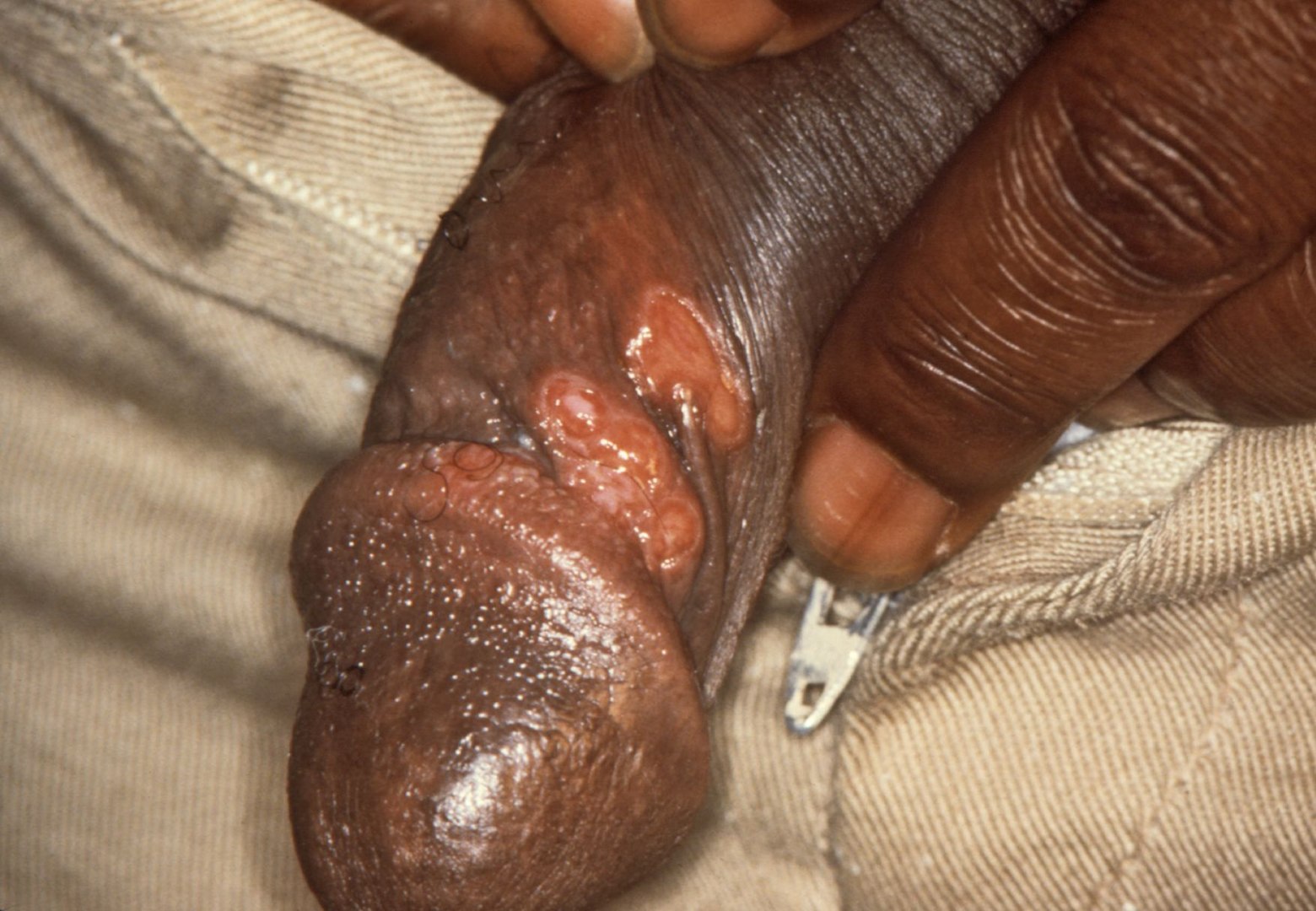
00:01 So let's talk about the clinical features of syphilis, and there are many. 00:07 First, primary syphilis. 00:09 At the sight of inoculation, we start out with a flat lesion called a macule which quickly develops into an elevated lesion called a papule, and then into a painless ulcer or chancre. 00:29 In men, this chancre will appear on the prepuce, the coronal sulcus, the shaft the corona and the glans in descending order of frequency. 00:41 Here are some examples of chancres on the glans, shaft, and coronal sulcus the prepuce and the labia in women. 00:53 This ulcer, one would think, would be painful but its actually painless and non-tender. 01:01 It may be that the spirochetes infiltrate the sensory nerves and that's why it's not painful. 01:11 And so lesions that are non-painful are the most characteristic. 01:20 The one exception would be a chancre in the anal area is generally very painful. 01:27 In patients who have HIV infection they may have actually multiple chancres. 01:34 Because they're not symptomatic, they may not even be recognized by the person who's been infected. 01:42 It may heal without recognition within 3-6 weeks. 01:49 That's when we find the development of secondary syphilis. 01:54 Secondary syphilis is the reason that this organism has been called the great imitator. 02:01 Because it can appear in a wide variety of anatomical locations and mimic lots of diseases. 02:10 But the bottomline is that the skin and mucous membranes are affected in 95% of individuals with syphilis. 02:21 They have rashes. 02:25 The characteristic rash -- and you can take this to the bank -- is a papulosquamous eruption, meaning that it's an elevated lesion that is scaly. 02:38 That is what papulosquamous means. 02:42 However, the lesions can be macular or flat, they can be papular, annular, pustular, or the worst form, Lues maligna, which is demonstrated in the lower panel, which is essentially an ulcerative form of pustular secondary syphilis. 03:08 So more about secondary syphilis. 03:11 Sometimes you can see lesions on the mucosa such as the oral mucosa or the labia. 03:22 And then we can find things that are determined Condylomata lata. 03:31 Now Condylomata acuminata are venereal warts, which is a subject for another presentation. 03:39 Condylomata lata are syphilitic and they are very highly contagious, teeming with organisms. 03:48 Now, these are elevated, sort of exophytic lesions, as you can see in these panels. 03:58 And at the same time, the patients usually have systemic symptoms. 04:01 They have low-grade fever and malaise. 04:04 They have arthralgias, myalgias, and generalized lymphadenopathy. 04:12 There's a real art to detecting axillary lymph nodes. 04:16 And in my experience, axillary lymphadenopathy is really quite common in syphilis and in an HIV infection. 04:25 Secondary syphilis can also involve the mucosa of the GI tract. 04:30 For example, it can heavily infiltrate the stomach, causing the stomach to be thick with or without ulcers. 04:39 And it can be mistaken for a stomach cancer. 04:45 The patients will often complain of epigastric pain and early satiety. 04:51 Now, the early satiety can be explained because the normal stomach when food comes in, stretches. 05:00 Whereas a thick wall of a stomach doesn't handle food as it comes in and the person who's affected may feel like they fill up too fast. 05:12 Or they may have frank nausea and vomiting. 05:16 When secondary syphilis involves the liver, you would expect an increase in the liver-associated enzyme especially the alkaline phosphatase. 05:27 But symphtomatic hepatitis is pretty rare in secondary syphilis. 05:34 Unfortunately, the eye can be involved and all structures from anterior to posterior may be involved. 05:44 The most characteristic is something called interstitial keratitis, which involves scarring of the cornea. 05:54 But the infection may be in the anterior chamber and cause an iritis or anterior uveitis. 06:02 It can be in the vitreous or choroid and cause a posterior uveitis or chorioretinitis, or an optic neuritis. 06:12 For example, the optic disc in optic neuritis becomes stark white. 06:21 The normal color of the optic disc is sort of bone-colored. 06:27 And if you were to look with an ophthalmoscope and saw that the optic disc was stark white, you've made the diagnosis of optic neuritis. 06:41 The ear can also be involved with hearing loss of a sensorineural nature. 06:49 The patient may complain of tinnitus or ringing in the ears or frank vertigo if the utricle and saccule are involved. 06:59 Worst of all, the brain can be affected. 07:04 And in secondary syphilis, you can actually have a syndrome that looks like meningitis or a syndrome like a stroke. 07:14 The bones can be involved and the classic is periostitis -- the outer surface of the bone. 07:22 The tibia is the most frequent bone affected by syphilis, and there is an example of syphilitic periostitis, the so-called saber shins. 07:36 But the sternum, the skull, and the ribs can also be affected in secondary syphilis.
About the Lecture
The lecture Primary and Secondary Syphilis by John Fisher, MD is from the course Genital and Sexually Transmitted Infections. It contains the following chapters:
- Primary Syphilis
- Secondary Syphilis
Included Quiz Questions
Which of the following best describes the classic rash of secondary syphilis?
- A diffuse, symmetric maculopapular rash involving the trunk and extremities
- A diffuse, symmetric vesicular rash involving the trunk and extremities
- A diffuse, symmetric maculopapular rash mainly involving the extremities
- A diffuse, symmetric maculopapular rash mainly involving the trunk
- A diffuse, symmetric vesicular rash mainly involving the trunk
Which of the following best describes a chancre?
- A 1-2 cm painless ulcer with a raised, indurated margin and a non-exudative base often associated with bilateral regional lymphadenopathy
- A 4-5 cm painless ulcer with a raised, indurated margin and an exudative base often associated with bilateral regional lymphadenopathy
- A 1-2 cm painful ulcer with a raised, indurated margin and a non-exudative base without regional lymphadenopathy
- A 4-5 cm painful ulcer with a raised, indurated margin and an exudative base without regional lymphadenopathy
- A 1-2 cm painless ulcer with poorly defined margins and an exudative base often associated with bilateral regional lymphadenopathy
What is the most common clinical manifestations of primary syphilis in immunocompetent patients?
- Painless ulcer at site of inoculation
- Localized tender lymphadenopathy
- Intensely pruritic vesicular lesions at the site of inoculation
- Dysuria
- Urethral discharge
Which of the following is NOT a sign of secondary syphilis?
- Condylomata acuminata
- Condylomata lata
- Generalized lymphadenopathy
- Fever
- Arthralgias
Periostitis of secondary syphilis is LEAST likely to affect which of the following sites?
- Femoral head
- Tibia
- Sternum
- Skull
- Ribs
Which of the following is the most common ocular manifestation of secondary syphilis?
- Uveitis
- Iritis
- Optic neuritis
- Dislocation of lens
- Retinal detachment
Which of the following sites/organs is generally NOT involved in secondary syphilis?
- Aorta
- Intestine
- Bone
- Skin
- Kidney
Author of lecture Primary and Secondary Syphilis

John Fisher, MD
Customer reviews
5,0 of 5 stars
| 5 Stars |
|
1 |
| 4 Stars |
|
0 |
| 3 Stars |
|
0 |
| 2 Stars |
|
0 |
| 1 Star |
|
0 |
Dr. Fisher is very thorough in explaining certain details but also very direct to point out important information.
