Playlist
Show Playlist
Hide Playlist
Portal-systemic Anastomoses

-
Slides Portal-systemic Anastomoses.pdf
-
Download Lecture Overview
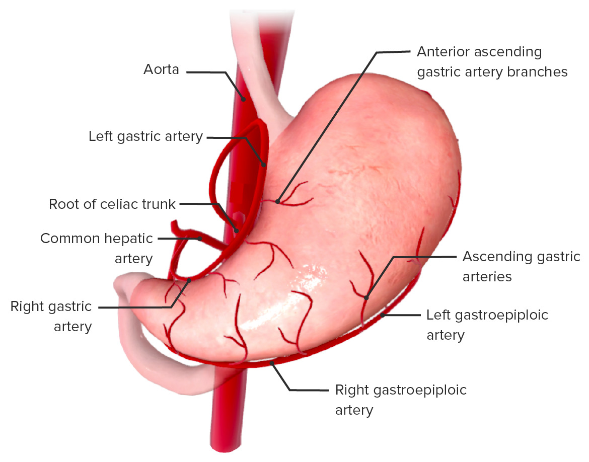
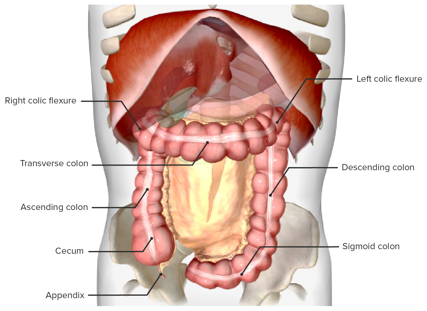
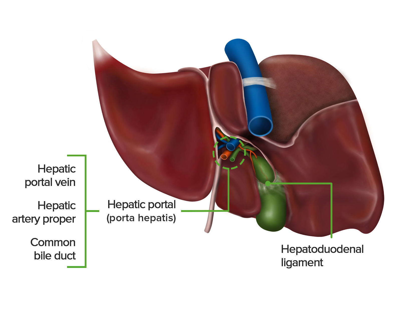
00:01 Now let's talk about the portosystemic anastomosis. 00:04 So what are these? These are anastomosis like we'd be familiar with before. 00:10 But instead of two arteries uniting together and forming those anastomosis coming from two different origins, this is going to be an anastomosis between the portal system and the systemic circulation of the body. 00:25 So the systemic portal anastomosis, these are going to be unions between the portal system and the general venous return of the body. 00:33 And they happen typically around four different places. 00:37 Why do they happen though? This is the normal blood flow, the venous return. 00:42 Superior mesenteric vein is going to be bringing blood oxygen low nutrient rich into the liver. 00:49 Alongside the inferior mesenteric vein which we see here draining into the splenic vein. 00:54 So here we can see the formation of the hepatic portal vein, which is now carrying all of that nutrient rich blood after the food has been ingested from the stomach and small intestines. 01:05 The hepatic portal vein then takes this nutrient rich blood into the liver where deamination for example can occur before that blood is then returned to the systemic circulation via the hepatic vein. 01:17 But what we can see is if we have liver cirrhosis, tumors, or general liver disease. 01:23 The flow of blood through the liver can be compromised. 01:27 Because the normal architecture of the blood vessels within the liver can become damaged. 01:33 We don't get the normal flow. 01:36 As we have limited flow going through the liver that can build up the blood pressure within this system. 01:42 That means the blood has to find alternative routes via the portal system and the general systemic circulation. 01:51 So let's have a look at a few of these. 01:53 So as we've seen, we've got esophageal and gastric veins. 01:57 We have paraumbilical and epigastric veins, We have colic and retroperitoneal veins. 02:03 And we also have some rectal veins at the bottom as well. 02:07 So let's start with the esophageal and gastric reins. 02:10 So here we see the esophageal and the gastric veins supplied around this junction between the esophagus and the stomach. 02:18 So here we have the gastroesophageal junction. 02:21 We can see the left gastric vein is giving rise to esophageal veins. 02:25 And here, we can have anastomosis with the azygos and hemiazygos venous supply. 02:32 So these esophageal veins will then actually join and form an anastomosis with both the azygos and the hemizygos system of veins which we can see here. 02:41 Now, the problem with this is that these veins are typically very small because they don't usually have that much blood of a high pressure and high volume passing through them. 02:51 And because that they can expand and lead to esophageal varices. 02:56 And here we can see them in the image. 02:58 These are expansions of these venous blood vessels passing into the lumen of the esophagus. 03:03 They can likely rupture and bleed out into the esophagus, and the patient can often cough up blood. 03:09 Let's have a look at a second one of these, and this is between the around the rectum. 03:14 Here we're gonna see the terminal branches of the superior rectal vein. 03:18 These are going to pass into the inferior mesenteric vein. 03:22 What we also have here is the middle and inferior rectal veins. 03:26 And again, these will be draining some of the blood away from the bottom portion of the rectum. 03:31 But if there's no increased blood pressure within the portal system, this can lead to increased blood now passing through the middle and inferior rectal veins, which will pass into the internal iliac vein. 03:44 The problem here is very similar to that of the esophageal varices. 03:49 Except this time, we call them hemorrhoids. 03:52 And these were the expansion of those blood vessels in the rectum can then bulge out of the rectum. 03:58 And these again can cause lots of bleeding, especially upon defecation. 04:03 Let's then have a look at the final two. 04:05 Here we have the paraumbilical and epigastric veins. 04:09 Now, this can lead to a whole series of enlarged venous structures along the anterior abdominal wall, as blood additional blood is now passing through inferior epigastric veins. 04:21 So here we can see the umbilicus. 04:23 And remember that the umbilicus and these power umbilical veins are really important, as they used to be embryologically quite active, quite open, allowing blood to pass through this region. 04:33 Within this region, we have these paraumbilical veins which are helping to drain blood from this region. 04:39 Now, with increased blood pressure around here, blood can now be forced into the superficial veins alongside the abdominal wall. 04:47 And this will feed into the inferior epigastric veins. 04:51 We've seen the inferior epigastric blood vessels which lie very superficial on the abdominal wall. 04:56 The veins we're running alongside them, and the same process happens here. 05:00 Blood wouldn't typically be running in these veins because they're very small. 05:04 And now we end up with these very large undulations on the anterior surface of the abdomen where the blood is now accumulated in these regions, where it typically wouldn't be found. 05:16 The final one is between here. 05:18 The colic veins which are draining the desending sigmoid aspects of the colon, and retroperitoneal veins. 05:25 These are veins which are draining the posterior abdominal wall. 05:28 And again, blood can now find a route back to the systemic circulation by utilizing this pathway. 05:35 And here we have colic veins anastomosing with retroperitoneal veins to form the four of these portosystemic anastomosis.
About the Lecture
The lecture Portal-systemic Anastomoses by James Pickering, PhD is from the course Vascular Supply of the Abdomen.
Included Quiz Questions
Portal-systemic anastomoses can be characterized as...? (Select all that apply)
- ...veins that connect the venous portal system to the systemic circulation.
- ...veins that typically carry large amounts of blood.
- ...veins that deliver additional blood to the liver when the portal circulation is blocked.
- ...veins that include the esophageal, rectal, para-umbilical, and colic veins.
- ...veins that are typically very narrow and quiescent.
Author of lecture Portal-systemic Anastomoses

James Pickering, PhD
Customer reviews
5,0 of 5 stars
| 5 Stars |
|
5 |
| 4 Stars |
|
0 |
| 3 Stars |
|
0 |
| 2 Stars |
|
0 |
| 1 Star |
|
0 |
