Playlist
Show Playlist
Hide Playlist
Plantar Aspect (Sole) of the Foot

-
Slide Plantar Aspect Foot.pdf
-
Download Lecture Overview
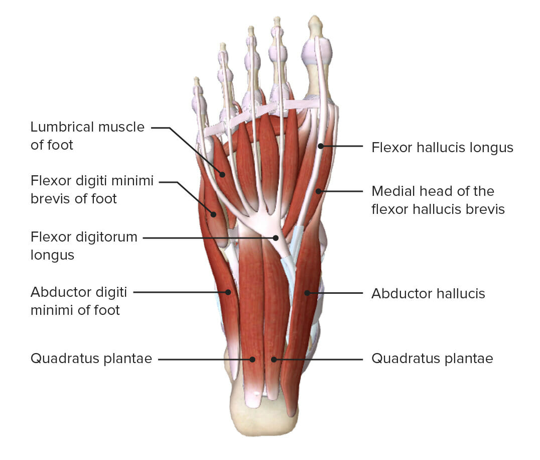
00:01 Now let's turn our attention to the plantar aspects of the foot or the sole of the foot, that inferior surface. 00:09 First of all, let's just discuss briefly the skin. 00:11 Because the skin on the sole of the foot is incredibly tough. 00:14 We have the heel pad here, most posteriorly on the sole of the foot. 00:19 We also have the ball of the great toe, which is quite a prominent landmark on this inferior surface. 00:24 And then we have the ball of the little toe, which is more laterally on this inferior surface. 00:29 These are toughened areas of skin as they bear a lot of pressure, a lot of weight as we walk, run, etc., during our daily movement. 00:38 If we were to remove the skin, then we'd see a very similar arrangement in what we see in the palm of the hand. 00:44 We have deep plantar fascia, which we can see here. 00:47 And then centrally, we have the plantar aponeurosis. 00:50 This is a very thick and tough band of connective tissue. 00:54 It's divided into a series of bands that contain the flexor tendons of the digits. 01:00 We'll come to those in the next few slides. 01:02 But these bands pass towards each and individual digits as they move distally within the sole of the foot. 01:09 Here we can see the superficial transverse metatarsal ligament. 01:13 This is important as it helps to bind and hold all of these ligaments in close position, helping to hold the tendons in position as they move towards the digits and preventing them from splaying. 01:25 If we were to look at the compartments of the foot, so here we're looking at a cross section, we can see, we have this plantar fascia here, or the plantar aponeurosis. 01:35 And that extends both laterally and medially as the lateral and medial plantar septi. 01:41 We can see the lateral and the medial plantar septi here in green. 01:45 This creates that central compartment and we'll discuss many of the muscles that we find in this compartment over the next few slides. 01:53 Lateral to this we have a lateral compartment, and then medially, we have a medial compartment. 01:59 We also have an interosseous compartment that resides between the metatarsal of the foot. 02:05 Most dorsally and we spoke about the various muscles in the dorsal aspect of the foot, we have this dorsal compartment. 02:13 All of this foot, we can see is surrounded by nice tough connective tissue, that deep plantar fascia, we also have that extending onto the dorsal surface of the foot where it's then covered by skin. 02:26 So now let's have a look at the muscles that make up the sole of the foot. 02:30 And there's quite a lot of muscles here and they've been divided into four layers. 02:36 So you may want to stop the video and make some notes but take this very slowly as there are a series of muscles here which we need to go through. 02:43 Also check with your own learning objectives from your own curricula to see how much detail you really need to know about this. 02:50 Do you really need to know all of the origins and insertions of all of these muscles because there are a lot of muscles here. 02:56 So take it steady and work through these slowly. 03:01 Let's have a look then at layer 1. 03:03 Layer 1, we have a series of muscles and these lie most superficial. 03:07 So they lay closest to the skin. 03:10 From lateral aspect, we have abductor digiti minimi, we then have flexor digitorum brevis, and then we have abductor hallucis. 03:19 Again, the names of these muscles will help give away their function. 03:23 If we then move on to layer 2, we see we have quadratus plantea muscle located quite centrally. 03:30 And then passing towards the digits over the metatarsals, we have the lumbricals. 03:35 In layer 3, we have a series of muscles that are associated with the big great toe, hence hallucis, and also one muscle associated with the little toe, the fifth digit, we have flexor digit minimi brevis and then we have adductor hallucis and flexor hallucis brevis. 03:54 Again, the names will indicate the function of these muscles. 03:58 If we then move on to the fourth layer, we see we have a series of interossei muscles, both the dorsal and the plantar interossei muscles. 04:07 These are similar to those we found in the hand.
About the Lecture
The lecture Plantar Aspect (Sole) of the Foot by James Pickering, PhD is from the course Anatomy of the Foot.
Included Quiz Questions
What is part of the plantar aponeurosis on the sole of the foot?
- Flexor tendons
- Extensor tendons
- Deep plantar fascia
- Superficial plantar fascia
Which muscle is most superficial in the foot?
- Abductor digiti minimi
- Quadratus plantae
- Lumbricals
- Flexor digiti minimi brevis
- Dorsal interossei
Author of lecture Plantar Aspect (Sole) of the Foot

James Pickering, PhD
Customer reviews
5,0 of 5 stars
| 5 Stars |
|
5 |
| 4 Stars |
|
0 |
| 3 Stars |
|
0 |
| 2 Stars |
|
0 |
| 1 Star |
|
0 |
