Playlist
Show Playlist
Hide Playlist
Pathogenesis and Clinical Features – Rheumatic Heart Disease

-
Slides Valvular Hypertensive Heart Disease.pdf
-
Download Lecture Overview
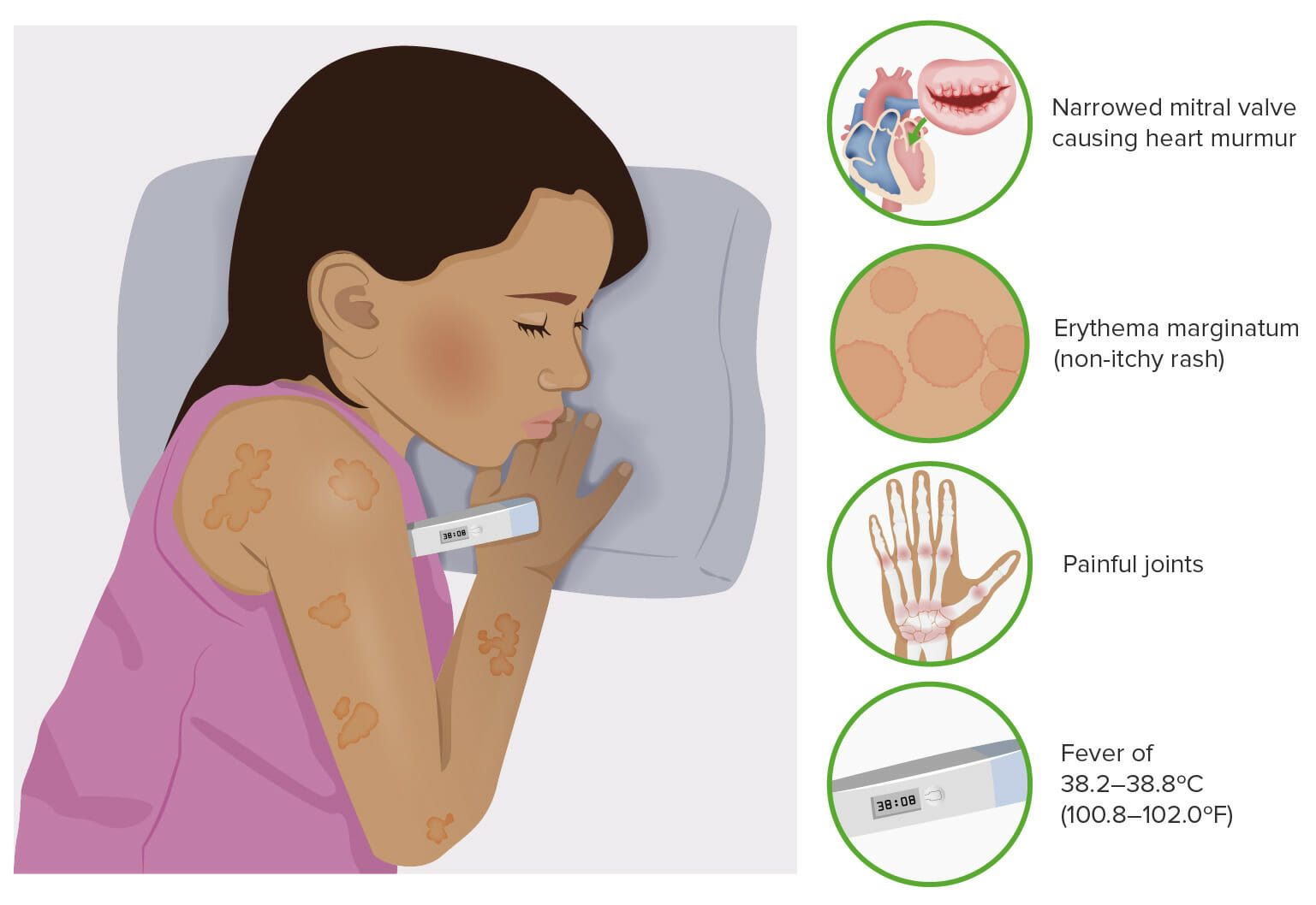
00:01 The exact pathogenesis is reasonably well understood. 00:06 So, here we have the Group A Streptococcus and on its surface are a number of antigens, proteins. 00:12 In particular, it's going to be the response to the M proteins on their capsule, on their surface that's going to be the major target. 00:22 When we develop an immune response, we have CD4 cells, plasma cells, antibodies, that are elicited that respond to the M proteins. And that's how we clear it. 00:31 The immune system effectively neutralizes the bacteria, binds antibody to them. 00:36 That will activate complement and also activate Fc receptor bearing cells and will get rid of the bug. The appropriate response. 00:44 However, depending on the strain of the streptococcus and depending on the unique antigenicity of any particular human host, there may be endothelial surface antigens or myocardial surface antigens or pericardial surface antigens that look just like those M proteins or look very similar to the M proteins. And here they are on a valve. 01:10 And now, in a patient who has developed the appropriate immune response to clear the infection, those antibodies bind to those similar antigens, and they are going to elicit the same complement activation, and they're going to elicit the same cellular response of FC receptor bearing cells and you're going to get tissue destruction as a result of that. 01:31 So, that's why you get this valvular disease. 01:34 What does it look like histologically? In the heart, this is what it looks like. 01:42 There's a very characteristic pathognomonic lesion called an Aschoff body or an Aschoff nodule. 01:50 And this is something that they do like to bring up periodically on the board, so pay attention. 01:56 In the myocyte, so you can also have not only just endothelial, valvular endothelial antigens that cross-react, you can have myocardial antigens that cross react. 02:08 And in that setting, you will get an accumulation of T lymphocytes, as well as some plasma cells, but basically, you're going to get very activated macrophages. 02:19 So, this is a DTH type response where the - delated type hypersensitivity response where the T lymphocytes that are recognizing this cross-reactive antigen on cardiac myocytes will activate macrophages, and they get a very characteristic look to them. 02:35 And they have a very abundant cytoplasm, but they have this slender ribbon of chromatin within their nucleus that makes the kind of their nucleus look like a little caterpillar has crawled through it. 02:47 That's the Anitschkow myocyte or the Anitschkow cell and it's a characteristic cell type within an Aschoff body. 02:55 And this is just basically a little focal delay type hypersensitivity response to cardiac antigens within the myocardium proper. 03:04 As a result of the macrophage activation, we get fibrinoid necrosis. 03:08 So, we get fibrin deposition, and we get killing of the myocytes. 03:11 This can actually be an acute cause of death where you can have sufficiently high numbers of these that you get arrhythmias. 03:19 The Aschoff bodies can be found anywhere within the three layers of the heart, and in fact, pancarditis, involvement of the endocardium, the myocardium, or the pericardium can all be seen in rheumatic heart disease. 03:34 So, we can find those Aschoff nodules on the endocardium and we would call it an endocarditis. 03:40 It's shown here on the endocardium of the left ventricle, but it can also be the endocardium of the valve. 03:45 If it involves the myocardium per se, it's myocarditis. 03:49 And if it involves the pericardium, it's called pericarditis. 03:53 And altogether, that's where we get the term pancarditis. 03:56 So, it - in any particular patient, you need not have all three of these areas in the heart involved. You can have one or more. 04:04 What does this look like? So, in acute rheumatic heart disease, the initial immune complex deposition plus/minus that T cell response leaves the little, tiny verrucae, small vegetations such as the verrucae are on the surface of the valve. 04:22 With time, as we recruit inflammatory cells, we will get scarring - we will get damage, and then we will get scarring. So, the leaflets thicken, and in many cases, will fuse at their commissures where the leaflets come together. 04:37 You can also get thickening and fusion of the chordae tendineae which will then leave the valve to become somewhat incompetent as that thickens. 04:45 And the classic lesion that we see is calcification bridging fibrosis across the valvular commissures. 04:53 So, instead of having the valve, watch my mouth, that opens widely, that's normally how the mitral valve opens. 04:59 Now, you have a valve [sound effect] that tends to be more like a fish mouth because the commissures are fused, and you don't open the leaflets as much. 05:10 And that's a great way to kind of think about the stenosis that occurs with rheumatic heart disease. 05:15 So, we have the mitral stenosis, thickening of the valves. 05:20 You have progressive dilation of the left atrium. 05:23 That's going to make it more prone to having atrial fibrillation, but also more prone to having retrograde flow into the pulmonary veins causing increased pressure throughout the lung and congestive heart failure. 05:37 The abnormal configuration of the left atrium also leads to potential thrombus formation. 05:44 We have abnormal flow on the left atrium. So, that can embolize. 05:47 We talked about having that emboli go to the brain or to a coronary artery or to some other part of the body. You can also have, with that progressive dilation left atrium, again, pressures moving retrograde through the lung, and then being reflected eventually in the right ventricle so you can have right ventricular failure and hypertrophy. 06:07 In general, the left ventricle is largely unaffected. 06:10 And having said that mitral rheumatic heart disease, it most commonly affects the mitral valve, secondarily affects the aortic valve, the tricuspid and pulmonic valves are rarely affected in rheumatic heart disease. 06:25 Pulmonic, almost never, rheumatic valvular disease in the tricuspid valve, perhaps 2 to 3% of cases. 06:33 And then, the aortic valve is affected 20 to 30% of the time. 06:37 So, kind of general clinical features that you need to be aware of. 06:41 You can have acute rheumatic fever, and onset of some of the valvular lesions or the pancarditis within 10 days to up to a couple months, and after group A streptococcal infection. 06:54 And notably, that number right there is very important. 06:56 The vast majority of people who even get a rip-roar in strep pharyngitis will not develop rheumatic heart disease. It only occurs in about 3% of patients. 07:05 It has to do, again, with the particular bug, its antigen, and its M protein, as well as the human host and its response to that M protein and how cross-reactive its own antigens are. 07:19 It will most often affect children, the acute rheumatic fever, in ages 5 to 15. 07:26 However, the manifestations of mitral stenosis take a long time to develop, and it's usually kind of mid to later life that you get significant enough damage to the mitral valve to cause the other secondary manifestations including left atrium thrombus, et cetera. 07:44 So, after the initial attack, you have an increased vulnerability to reactivation. 07:51 So, every time now a patient who's had strep pharyngitis with rheumatic heart disease, the next time they get a strep infection, they already have a slightly abnormal valve, and they already have a relatively high titer of antibodies and T cells that can respond. 08:06 So, each time they get another strep pharyngitis, they are more likely to compound the initial injury and make it worst and worst and worst with each subsequent infection.
About the Lecture
The lecture Pathogenesis and Clinical Features – Rheumatic Heart Disease by Richard Mitchell, MD, PhD is from the course Valvular and Hypertensive Heart Disease.
Included Quiz Questions
What aspect of the mitral valve is at risk of being attacked by the immune system in rheumatic heart disease, and why?
- Endothelial surface antigens, due to their similarity to bacterial M proteins
- Papillary muscle fibers, due to their similarity to bacterial surface proteins
- Mitral annulus cells, due to a higher likelihood of calcification
- Mitral valve cusp cells, due to a higher likelihood of bacterial deposition
- Endothelial surface antigens, due to a higher likelihood of cell damage
Which of the following histological features is NOT identified within an Aschoff body?
- Caseous necrosis
- T lymphocytes
- Anitschkow cells
- Fibrinoid necrosis
- Plasma cells
Calcification of what structure(s) creates the “fish mouth” appearance of the mitral valve in rheumatic heart disease?
- Valvular commissures
- Papillary muscles
- Valvular cusps
- Chordae tendineae
- Verrucae
Which of the following structures is typically unaffected by rheumatic heart disease?
- Left ventricle
- Mitral valve
- Right ventricle
- Left atrium
- Aortic valve
What percentage of patients who contract group A streptococcal infection develop rheumatic heart disease?
- 3%
- 10%
- 0.5%
- 25%
- 40%
When do patients typically manifest symptoms of acute rheumatic fever and rheumatic heart disease, respectively?
- Fever: ages 5–15 Heart disease: mid-to-later life (ages 40–60)
- Fever: ages 1–5 Heart disease: adolescence (ages 15–20)
- Fever: ages 25–40 Heart disease: elderly (60+)
- Fever: ages 5–15 Heart disease: early adulthood (ages 20–30)
- Fever: ages 30–40 Heart disease: mid-to-later life (ages 40–60)
Author of lecture Pathogenesis and Clinical Features – Rheumatic Heart Disease

Richard Mitchell, MD, PhD
Customer reviews
5,0 of 5 stars
| 5 Stars |
|
1 |
| 4 Stars |
|
0 |
| 3 Stars |
|
0 |
| 2 Stars |
|
0 |
| 1 Star |
|
0 |
Dr. Mitchell is awesome! The fish mouth description made me laugh but I will never forget what it means lol. Thank you!
