Playlist
Show Playlist
Hide Playlist
Obstructive Uropathy (Post-renal AKI) with Case

-
Slides Nephrology Acute Kidney Injury.pdf
-
Download Lecture Overview
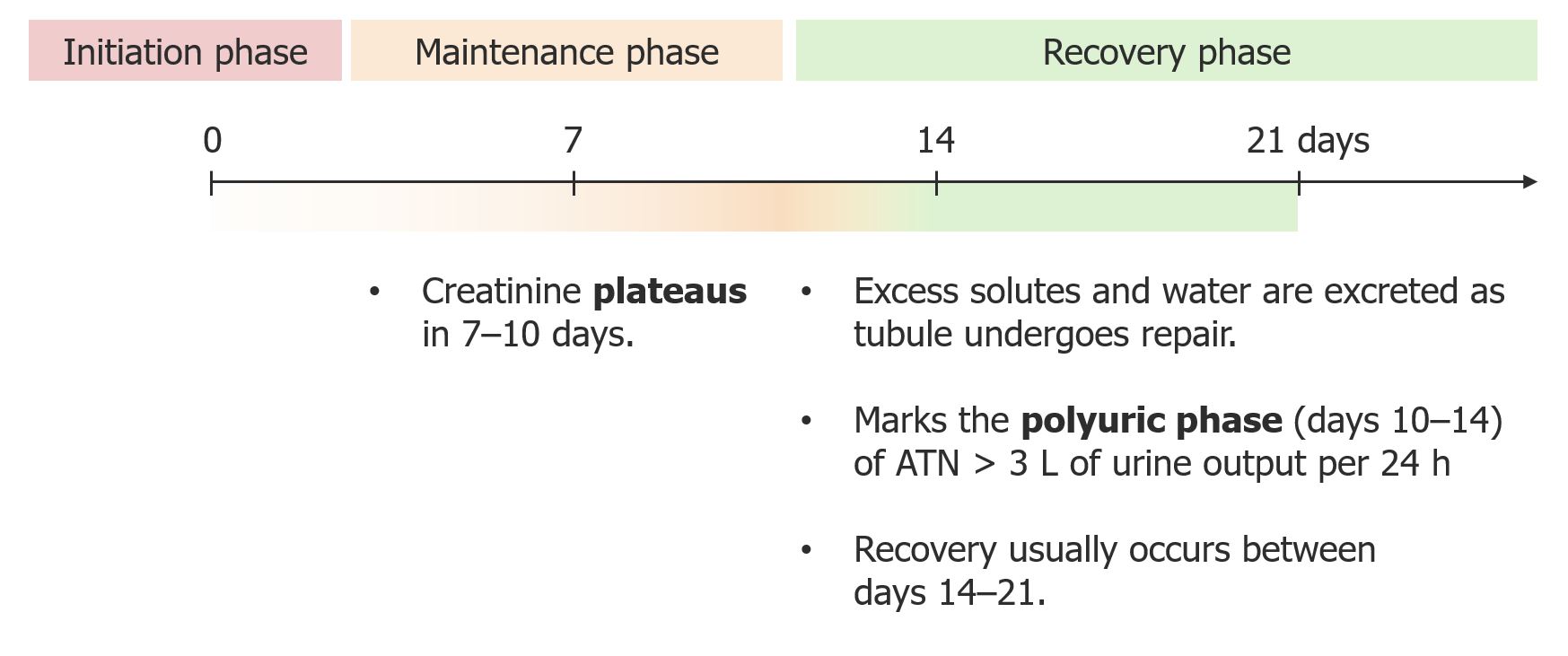
00:01 So this kind of brings us to our very last category of the categories of AKI - urinary obstruction. 00:08 So this really is going to be caused by obstruction of the outflow of urine anywhere from the renal pelvis to the urethra. 00:17 And the renal failure that we see caused by this can either be acute or it can be subacute if it occurs over time. 00:25 When we think about the different causes of obstructive uropathy, they include calculi or stones particularly if I have a stone or a patient has a stone at the ureteropelvic junction in a solitary kidney obstructing outflow of urine that's gonna result in AKI. 00:42 Or think about a patient who might have bilateral stones And not only do they have little stones, they have these big staghorn calculi that take up the entire renal pelvis, that can really obstruct the outflow of urine So that can result in an AKI. 00:58 And when we have stone disease, we can see it in younger patients as well as older patients. 01:04 We can also have anatomic abnormalities most commonly seen in children. 01:09 So children who have problems with the positioning of the urethral valves, people who are born with congenital strictures or adults who develop that over time because of a traumatic injury, and then people who have stenosis of the ureteral vesicle or the ureteropelvic junction Again, that just means that there's an area of narrowing where the ureter anastomoses with the bladder or the ureter is connected to the renal pelvis. 01:36 Now the most common and what we saw in our case is benign prostatic hyperplasia or BPH. 01:42 This is most common in men over the age of 50. 01:47 Another is urethral stricture as we talked about. 01:51 And then finally, malignancy. 01:53 So malignancy which will include malignancies of the prostate, the bladder or any kind of extrarenal pelvic neoplasms that end up compressing the outflow of urine essentially from those ureters. 02:06 Okay, so if our patients presents with obstructive uropathy, what can we do in order to make that diagnosis? Again, we're gonna be diligent detectives, we're gonna do a very thorough history and chart review. 02:17 If our patient has BPH, we're really gonna ask them about lower urinary tract symptoms. 02:22 That means do they have hesitancy when they're voiding? Do they have dribbling? Do they have double voiding, feeling like they've voided, but then have to void again? That means that they have incomplete emptying and that's a very big sign or symptoms that this patient has BPH. 02:37 In a patient who might be presenting with stones, they could present with flank pain depending on where that stone is located, or they can have gross hematuria, again as that stone kind of traverses that ureter or in the bladder even you can develop hematuria. 02:54 And then of course, for people who have malignancies that are compressing the ureters then it's gonna be important to look for that history of malignancy. 03:03 Physical exam can be helpful as well in terms of diagnosing our patients. 03:08 For BPH, it would be important to do a digital rectal exam to palpate for an enlarged prostate, In patients who have stones, we might want to do percussion at our costovertebral angle to see if our patient has tenderness. 03:23 And then in the case of malignancy, it's always important to do a thorough abdominal exam to really palpate for any abdominal mass in the pelvic and abdominal region. 03:34 Imaging is probably one of the most important parts in diagnosing post-renal or obstructive uropathy. 03:41 And our imaging modality of choice is going to be renal ultrasound. 03:45 Here we can look for things like hydronephrosis. 03:48 As seen in the image over here, you can see on ultrasound we have a kidney with a very dilated renal pelvis and a very big boggy renal cortex. 03:58 That's from essentially reflux of urine back into that kidney, enlarging it. 04:04 If our patient is presenting with a stone, it'd be important to get a CT of the abdomen and pelvis without radiocontrast which will highlight the stone - so very easy to see calculi. 04:14 We can also see pelvic masses if we want to use contrast although we'd want to avoid that in our patients who have AKI. 04:20 On laboratory evaluation, that high BUN to creatinine ratio is gonna be more reflective of post-renal. 04:28 Again, if you think about what's happening, you have obstruction of urine outflow, so you have stasis of urine within that tubule so plenty of opportunity to absorb urea during that time period. 04:39 And again if I'm looking at that urinary sediment, it's oftentimes bland. 04:43 There's no cellular elements, there's no cast. 04:46 In the case of nephrolithiasis or stones, I may be lucky enough to see some crystals but they're not always present. 04:54 So how do we treat our patients once they present with obstructive uropathy? So for BPH, we can actually do a urinary catheter inserted into the bladder for decompression We can also remove medications that precipitated the obstruction to begin with. 05:12 Remember our patient, he took a cold medication which was an alpha-agonist. 05:16 So that increases tone of that bladder. 05:18 Next we absolutely want to ensure our patients are off-medications that it can do things like that. 05:23 And there's also medical and surgical therapy for prostate. 05:27 Right, so we're gonna give your patients alpha-blockers. 05:30 And we can have our urology colleagues get involved and they can definitively cure our patients with BPH. 05:38 For stones, we can remove the stone and that's when I typically get my urology colleague involved where they can do a percutaneous nephrolithotomy or they can do shockwave lithotripsy. 05:50 We'll be talking a little bit more about that in our nephrolithiasis chapter. 05:55 We can also have our urology colleagues place a ureteral stent from that renal pelvis all the way to the bladder which helps to keep that ureter open and allows free passage of stones into the urine. 06:05 And finally, we can get our interventional radiology colleagues involved to place a percutaneous nephrostomy as shown in this image over here. 06:13 So essentially, our IR colleagues will place the catheter directly into the renal pelvis which completely obviates the obstruction and allows free passage of urine.
About the Lecture
The lecture Obstructive Uropathy (Post-renal AKI) with Case by Amy Sussman, MD is from the course Acute Kidney Injury (AKI).
Included Quiz Questions
Which of the following is NOT a common cause of obstructive uropathy in children?
- Horseshoe kidney
- Urethral valves
- Strictures
- Stenosis at the ureterovesical or ureteropelvic junction
Which of the following is the most likely symptom of benign prostatic hyperplasia?
- The feeling of incomplete voiding
- Pain on urination
- Hematuria
- Milky penile discharge
Which imaging test is best for identifying calculi?
- Abdominal computed tomography without contrast
- Abdominal computed tomography with contrast
- Ultrasonography of the kidney
- Abdominal X-ray
Author of lecture Obstructive Uropathy (Post-renal AKI) with Case

Amy Sussman, MD
Customer reviews
5,0 of 5 stars
| 5 Stars |
|
5 |
| 4 Stars |
|
0 |
| 3 Stars |
|
0 |
| 2 Stars |
|
0 |
| 1 Star |
|
0 |
