Playlist
Show Playlist
Hide Playlist
Neurovasculature of the Large Intestine

-
Slides Neurovasculature of the Large Intestine.pdf
-
Download Lecture Overview
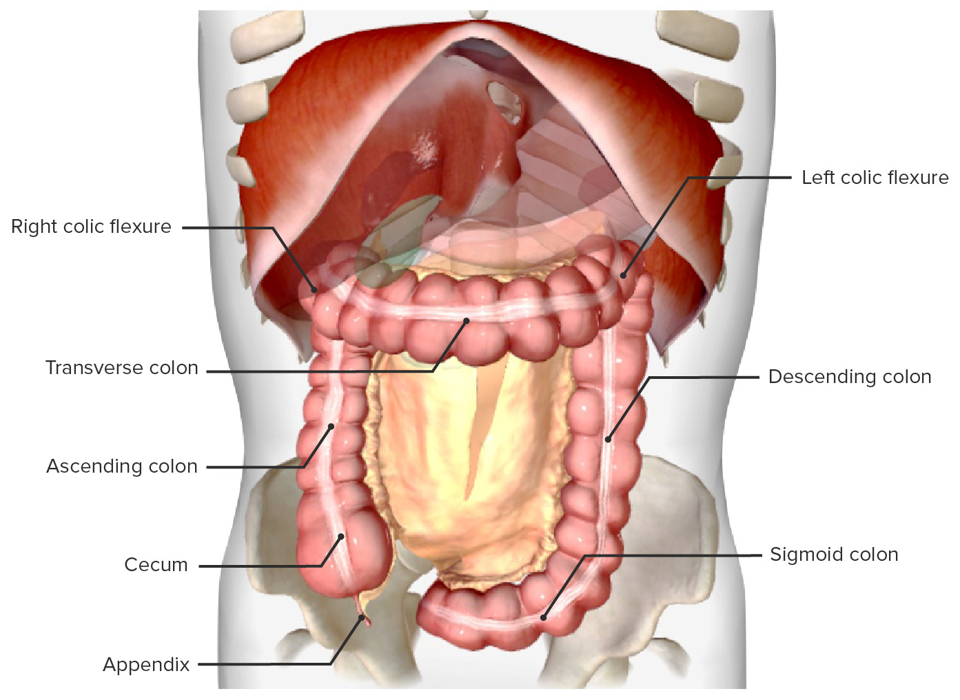
00:01 So let's just move on to the vasculature of the large intestine and see how the large intestine is supplied. The large intestine is an interesting organ because it is really a derivative of both the mid gut and the hind gut. 00:14 And if you remember, the mid gut and the hind gut are going to be supplied by the superior mesenteric artery for the mid gut and the inferior mesenteric artery for the hind gut. 00:24 So, one continuous chew but actually there's the mid gut and the hind gut, so we have 2 blood vessels that go and supply it. It's important to note the transition between this fold by the marginal artery, and we'll come to that. So here we can see represented on the screen, we have the abdominal aorta and here we can have the superior mesenteric artery that's running down. And here we can also see the inferior mesenteric artery. If we were to have a look at the superior mesenteric artery, then coming off it we have the ileocolic artery which is going to both bits of ileum, ileum and the colon with the colic branches. So we can see the ileocolic artery giving rise to both the ileal and the colic branches supplying both bits of the ileum, small intestine, and the cecum, the large intestine via the colic branch. We then have the right colic artery. We also have the middle colic artery. So all of these blood vessels coming away from the superior mesenteric artery. 01:23 If we then go across to the other side, we can see we have the left colic artery. And the left colic artery here is coming off the inferior mesenteric artery. So we can see the left colic, we can see the sigmoidal artery, and we can see the superior rectal artery here. And these are coming off the inferior mesenteric artery and it's important to appreciate now how we're really distinguishing between mid gut and hind gut. 01:48 The mid gut regions is supplied by the superior mesenteric artery whereas the hind gut is supplied by inferior mesenteric artery. But there is a very important feature that we need to be aware of and that is here between the left colic artery coming from the inferior mesenteric artery and the middle colic artery which is coming from the superior mesenteric artery. Now these essentially run towards the colon and then they form these arterial arcades that really run towards the actual chew bit self. So we got these kind of main highways that run towards them and then they form these arterial arcades that run along the inner side of the large intestine forming this kind of connecting loop. And this is an anastomotic loop, very similar to what we saw around the greater and lesser curvatures of the stomach. And this connecting loop between the middle colic and the left colic is known as the marginal artery. And that's really forming this anastomosis you can see here. Typically, it course most prominently in the upper left quadrant and this is where really you find the distinction between the midgut, the transverse colon's proximal 2/3, and the transverse colon's distal 3rd. 03:02 So the last 3rd of the transverse colon is hind gut predominantly supplied by inferior mesenteric whereas the proximal 2/3 of the transverse colon is mid gut predominantly supplied by the middle colic artery by the superior mesenteric artery. But where that leads the transition it's supplied by the marginal artery which runs and helps to supply this region. This is important because it builds in some redundancy into the system. 03:32 So if we were to have an inclusion and occlusion of the inferior mesenteric artery, very common site for the abdominal aorta to become blocked, then actually you'd think the hind gut wouldn't be able to have a blood supply. But by way of the marginal artery, blood from the superior mesenteric artery can feed into the hind gut and replace the blood supply of the inferior mesenteric artery. You can see the blue dotted lines here indicating that movement of blood. So now let's just briefly finish up by looking at the nerve supply to the large intestine. And again, it's important to appreciate that it's both the midgut and the hind gut structure. So we have inputs from both the superior mesenteric plexus and the inferior mesenteric plexus. The superior mesenteric plexus is going to give rise to the sympathetic and parasympathetic supply coming from both the vagal nerve and coming from the sympathetic chain and that's going to supply the mid gut. 04:27 Whereas the inferior mesenteric plexus is going to be receiving sympathetics from the pelvic region and also parasympathetics from the pelvic region as well and they're going to be supplying the hind gut. But the superior and inferior mesenteric plexus are the ultimate nerve hobs for both the mid gut and the hind gut.
About the Lecture
The lecture Neurovasculature of the Large Intestine by James Pickering, PhD is from the course Anatomy of the Large Intestine.
Included Quiz Questions
Which structure is a branch of the inferior mesenteric artery?
- Left colic artery
- Middle colic artery
- Right colic artery
- Ileocolic artery
- Inferior pancreaticoduodenal artery
At which vertebral level does the inferior mesenteric artery originate from the abdominal aorta?
- L3
- T10
- L5
- T12
- L1
An inferior mesenteric artery is occluded. Which artery can provide an alternative blood supply that can prevent ischemia of the descending colon, sigmoid colon, and rectum?
- Middle colic
- Gastroduodenal
- Right colic
- Splenic
- Ileocolic
Author of lecture Neurovasculature of the Large Intestine

James Pickering, PhD
Customer reviews
5,0 of 5 stars
| 5 Stars |
|
5 |
| 4 Stars |
|
0 |
| 3 Stars |
|
0 |
| 2 Stars |
|
0 |
| 1 Star |
|
0 |
