Playlist
Show Playlist
Hide Playlist
Neurovasculature of the Anterior Compartment of the Arm

-
Slide Neurovasculature of Anterior Compartment Arm.pdf
-
Download Lecture Overview
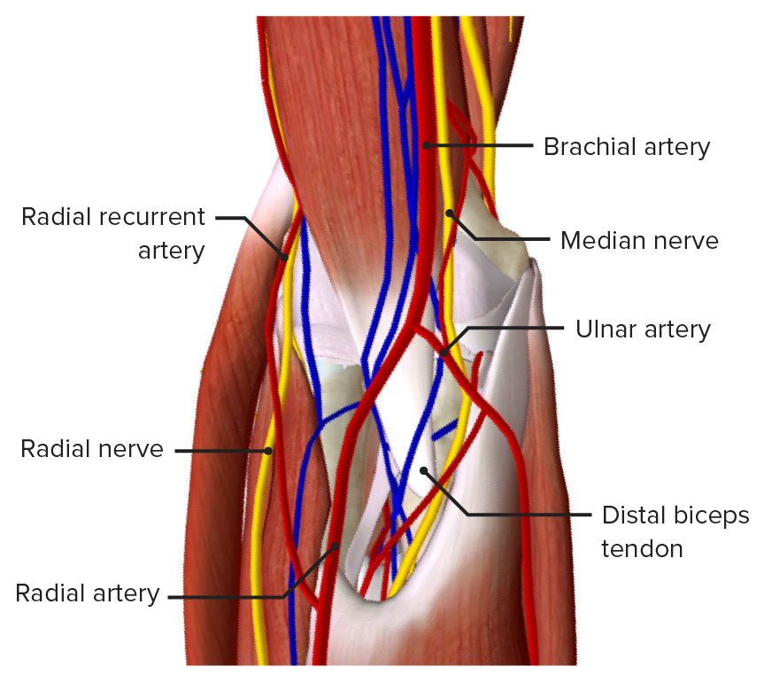
00:01 So, now, let's just have a look at some of these muscles kind of in situ and its relationship to other muscles. 00:06 So, we can see biceps brachii here in the anterior aspect. 00:10 A lot of its tendinous insertions kind of superiorly towards the clavicle and towards the scapula, forgive me, are actually covered by various muscles. Here, we can see pectoralis major and deltoid muscle really covering those tendinous insertion points around the shoulder joint. 00:26 If we remove the pectoralis major, we can see the short head. 00:29 If we remove deltoid muscle, we can see the long head. But you can see their relationship to those muscles. 00:36 If you remove brachialis muscle, then, deep to it, you see biceps brachii. 00:41 And then, most medially in this compartment, we see coracobrachialis. 00:45 Brachialis muscle can then, just be added on top as the most superficial muscle within this region. 00:52 The blood vessel that runs down alongside it really is the axillary artery but within the brachium, within the arm itself, it becomes the brachial artery and this happens at the lower border of teres major. 01:04 And the brachial artery is that important blood vessel where you take for blood pressure measurements. 01:08 And you can feel it. You can feel the brachial pulse if you find the cleft on the medial aspect of brachialis muscle. 01:15 The brachial artery then, continues along the medial aspects of brachialis muscle to enter the cubital fossa. 01:21 And here, we can see a midway point between the two epicondyles of the humerus, the lateral and the medial epicondyles. The brachial artery splits into its two braches that supply the forearm, the radial artery laterally, and the ulnar artery more medially. 01:39 We see these within the cubital fossa which we'll come to later on. 01:42 And these are sitting on top of the biceps tendon which we can see there. 01:47 Coming off the brachial artery, we can see the superior ulnar collateral artery and we can see its equivalent inferior version. So, these two are helping to supply muscles within this region and also, the joint capsule around the elbow. 02:02 If we look slightly more superiorly, then, coming off the brachial artery just as its formed, so, just as the axillary artery has ended leaving the axilla, we have the profunda brachii artery. 02:14 And this passes posteriorly through the triangular interval with the radial nerve to supply the posterior compartment of the arm. 02:22 And here, we can see it's being accompanied by that radial nerve. 02:27 The median nerve runs alongside the brachial artery. 02:30 Remember, the median nerve is coming away from the two aspects of the lateral and medial cord. 02:35 We can see it running in close association with the brachial artery here, running lateral to it. 02:41 And then, we have it running as it passes more inferiorly, medially to the brachial artery. 02:47 And this is an important relationship we'll come to see later on when we look at the cubital fossa. 02:53 Here, we can see running medial to the brachial artery, we have the ulnar nerve. 02:57 Remember, the ulnar nerve is the direct continuation of the medial cord of the brachial plexus. 03:03 So, we have some very important structures running from the axilla into the anterior compartment of the arm. 03:10 We mentioned one important gateway through to the posterior aspect when we looked at the triangular interval, radial nerve and profunda brachii artery. 03:19 We also have some branches thatpenetrate the kind of medial intermuscular septum that helps branches pass from the anterior compartment into the posterior compartment. 03:29 And here, we can see some blood vessels passing in that direction. 03:33 We can add on a couple of cutaneous branches so the medial, brachial, and antebrachial cutaneous nerves, respectively, supplying skin in those regions.
About the Lecture
The lecture Neurovasculature of the Anterior Compartment of the Arm by James Pickering, PhD is from the course Anatomy of the Arm.
Included Quiz Questions
Which muscle lies most medial and posterior to the biceps brachii muscle?
- Coracobrachialis
- Triceps brachii
- Deltoid
- Brachioradialis
- Teres major
What is the relationship of the median nerve to the brachial artery in the upper half of the arm?
- Lateral
- Medial
- Anterior
- Posterior
- Superior
Author of lecture Neurovasculature of the Anterior Compartment of the Arm

James Pickering, PhD
Customer reviews
5,0 of 5 stars
| 5 Stars |
|
5 |
| 4 Stars |
|
0 |
| 3 Stars |
|
0 |
| 2 Stars |
|
0 |
| 1 Star |
|
0 |
