Playlist
Show Playlist
Hide Playlist
Myocardial Infarct: Complications

-
Slides Complications of Atherosclerosis.pdf
-
Download Lecture Overview
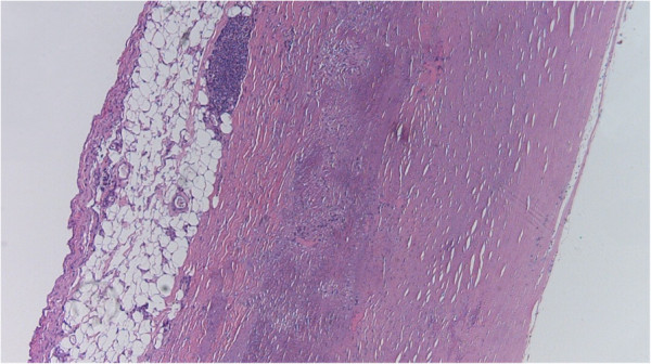
00:01 Let's talk about complications then. 00:03 And they're predictable based on everything that you've just learned, right? Okay, so here we have a transverse section of myocardium. 00:11 The right ventricles on the right, identified with RV, left ventricles on the left (LV), and we have with arrows pointing to an area of transmural, lateral wall myocardial infarction. 00:27 If we were in the classroom together, I'd say what vessel was occluded? Those of you who are paying attention at home, this is the left circumflex. 00:34 This is a beautiful left circumflex distribution. 00:37 And we have a transmural infarct, now some of this infarct, the area with a smaller arrow at the bottom is just showing you kind of what granulation tissue looks like. 00:49 It's a little bit glassy, it's a little bit depressed, and all that kind of yellow tan stuff in the middle, surrounded by that glassy granulation tissue is necrosis. 01:00 It's what necrotic myocardium looks like versus the healthy myocardium and the rest of the heart. 01:05 The big arrow pointing to that hemorrhagic thing, we're looking at a transmural, free wall rupture. 01:12 And so this patient was recovering nicely from their myocardial infarct. 01:18 And at about 7 to 10 days out as we got to maximum granulation tissue and maximal destruction of the entire thickness of the wall, a heart squeezed, and then ruptured laterally. 01:30 That blood that was in the ventricle now is in the pericardial sac. 01:34 As a consequence of that, the patient develops terminal tamponade. 01:39 The heart is continuing to squeeze, but it cannot fill any more because there's blood in the pericardium. 01:46 And that's, you're looking at the cause of death in this patient. 01:49 Another complication. 01:50 So it can't, it doesn't have to be necessarily a free wall rupture. 01:55 You can have rupture of the papillary muscle, for example. 01:58 and that's what's being demonstrated here shown here with the little skinny white arrow, is pointing to a papillary muscle that would normally be holding parts of the mitral valve which is shown that kind of arching, yellow thing at the top. 02:15 And it has now had a transmural infarct of the papillary muscle. 02:20 And as the heart continues to beat, that completely ruptures. 02:23 Now, we have wide open mitral regurgitation. 02:27 We have a flail leaflet because the papillary muscle is gone. 02:30 And this patient would have been going along quite nicely. 02:33 In about 7 to 10 days out, suddenly developed wide open mitral regurgitation, and florid fulminant pulmonary edema, couldn't breathe. 02:43 And as a result of that, that's why the patient died. 02:48 You don't have to have free wall, you don't have to have papillary muscle rupture, you can have the interventricular septal rupture. 02:54 That's what's being demonstrated here. 02:56 So the right ventricle on this cardiac echo is, actually I believe this is cardiac MRI. 03:04 That right ventricle is shown there with the RV, the LV is shown up top with an LV label, left ventricle and the arrows are pointing to an area between the left ventricle and the right ventricle where blood is now flowing from the left ventricle under higher pressure into the right ventricle. 03:20 So this is an inter ventricular septal rupture. 03:23 This will actually cause left to right shunting with acute pulmonary vessel vascular overload, can actually cause acute right heart failure, and that can also be a cause of death. 03:36 So predictably, because of the way that the heart heals after an infarct, you can predict some of the effects that can occur and why they occur when they occur. 03:49 And that's mainly because of the granulation tissue that's been generated as part of the healing process. 03:58 Let's say you live long enough. 04:00 Remember we talked about how there would be thinning and remodeling of the ventricle as scar is formed, and then it kind of recalibrates, it gets thinner as it remodels. 04:15 That remodeling of the chamber under pressure will lead to the formation of an aneurysm. 04:20 That's what we're demonstrating here. 04:22 The big white area with the arrow is a very dilated, left ventricle chamber. 04:31 You can see the normal thickness of the wall, which is what's present in the interventricular septum. 04:36 Kind of in the middle. 04:38 And then you can see that the lateral wall is markedly thin. 04:42 So we have this big globoid heart. 04:45 Keep in mind that all of that thin part of the wall is not going to be very contractile. 04:50 Now thinking about Virchow's triad, remember that abnormal flow. 04:56 And this is definitely going to be abnormal flow because it's not squeezing at all. 04:59 It's not moving. 05:00 We're going to form a thrombus and what the arrow is pointing to, at the apex of the heart is a thrombus that's occurred in this ventricular aneurysm. 05:09 So not only is the heart not squeezing because of the transmural infarct but they have abnormal flow and Virchow's triad is allowing us to form a thrombus that can potentially embolize. 05:21 And then when it embolizes, the next stop is going to be the brain or some other important structure.
About the Lecture
The lecture Myocardial Infarct: Complications by Richard Mitchell, MD, PhD is from the course Atherosclerosis.
Included Quiz Questions
What can happen after the complication of free wall rupture after a myocardial infarction (MI)?
- Terminal tamponade
- Compensatory hypertrophy
- Necrotic tissue formation
- Aneurysm
- Granulation tissue formation
Free wall rupture can cause blood to fill in the...
- ...pericardial sac.
- ...aortic aneurysm.
- ...auricles.
- ...septal tissue.
- ...ventricles.
Papillary muscle rupture can cause...
- ...mitral regurgitation.
- ...compensatory hypertrophy.
- ...ventricular aneurysm.
- ...scar tissue contracture.
- ...mitral stenosis.
Author of lecture Myocardial Infarct: Complications

Richard Mitchell, MD, PhD
Customer reviews
5,0 of 5 stars
| 5 Stars |
|
5 |
| 4 Stars |
|
0 |
| 3 Stars |
|
0 |
| 2 Stars |
|
0 |
| 1 Star |
|
0 |
