Playlist
Show Playlist
Hide Playlist
Myasthenia Gravis: Repetitive Stimulation – Diagnosis

-
Slides Strowd Myasthenia Gravis.pdf
-
Download Lecture Overview
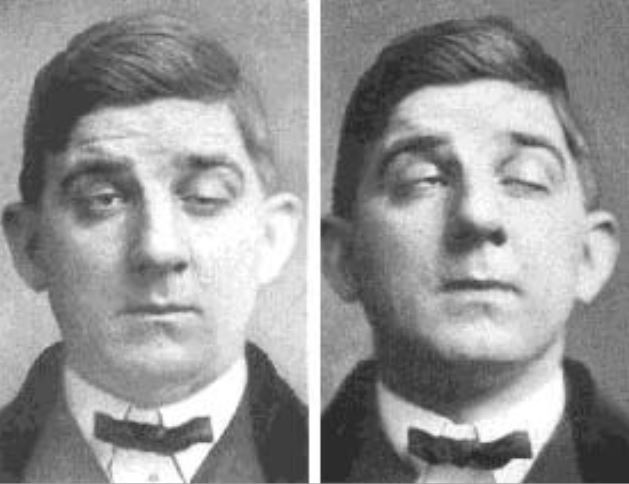
00:01 Let's look a little bit more closely at what's happening with repetitive stimulation in both a normal and myasthenic case. 00:08 What we see here on the top graph is how muscles work. 00:12 In order for a muscle to contract, there must be an end-plate potential. 00:17 And that's depolarization of the muscle that drives contraction. 00:21 In the normal state, if we activate the muscle, the end-plate potential is above this dashed line. 00:27 And that's the amount of end-plate potential needed for a muscle to contract. 00:31 And so when the nerve sends an action potential down the presynaptic terminus, there's enough acetylcholine quanta in the synaptic cleft to drive the muscle to contract. 00:42 If we activate the nerve again, activate that muscle again, we see that again, the signal is higher than that safety factor, and the muscle will contract again. 00:51 If we activate the nerve, again, the muscle will contract then again, and again, and again, the amount of acetylcholine quanta that are released in the synaptic cleft is sufficient to drive muscle contraction. 01:01 There is a little bit of a dip, but it always remains above this safety factor. 01:06 The amount of end-plate potential needed for the muscle to contract. 01:11 What happens in myasthenia gravis? When we see on the top graph, that the initial signal, the amount of end-plate potential that's generated, as a result of blocking of that postsynaptic signal is enough to generate muscle contraction. 01:25 It's over the safety factor, but not near as high as the normal condition. 01:29 And with repeated signaling with repeated action potentials, we see that the number of acetylcholine quanta that's in the synaptic cleft falls below the safety factor. 01:38 The muscle does not contract, and patients present with weakness. 01:42 What does that look like on the nerve conduction study? Well, here we're looking at what an electrophysiologist would be looking at. 01:50 When they shocked the nerve, they see the muscle contracts. 01:53 And each one of these curves is muscle contraction. 01:56 And if we activate the nerve and tell it to move the muscle, the muscle will contract with the first shock, with the second shock, the third, fourth, fifth, sixth, seventh, eighth shock as well. 02:06 And we always see that for each shock, there is muscle contraction. 02:11 In the myasthenic state, when there's blocking of that signal that transmission through the neuromuscular junction, we see with the first activation of the nerve, enough acetylcholine quanta is released into the synaptic cleft and the muscle moves. 02:25 But with repetitive stimulation with repeated activation of that nerve, there is less and less end-plate potential less and less acetylcholine quanta that activates that acetylcholine receptor, and the muscle contraction shrinks, and we see less contraction and a decremental response. 02:42 And that decremental response is diagnostic of a neuromuscular junction disorder and is seen in patients with myasthenia gravis. 02:52 So again, the normal process acetylcholine is stored in the presynaptic terminus in both primary, secondary, and tertiary stores. 02:59 That's how the nerve stores acetylcholine. 03:03 The primary store is freely available, but it is rapidly depleted. 03:07 And it is used with each of those stimulations that we saw in that repetitive stimulation diagram. 03:15 The secondary store is mobilized with continuous maximal exertion. 03:19 So if you activate a muscle continuously for one minute, you deplete all of the primary stores of acetylcholine, and you start to activate the secondary stores. 03:29 That's your mobilized store of acetylcholine. 03:33 And then there are tertiary stores that are reserved for special and rarely utilized situations. 03:41 A decremental response can be seen in any patient but should not be more than 10%. 03:46 And a decrement with slow repetitive nerve stimulation of more than 10% is diagnostic of a neuromuscular junction disorder. 03:54 And as seen in patients with myasthenia gravis. 03:57 This decrement typically occurs between three to five minutes. 04:02 So what's working? What's happening? What's going on at the neuromuscular junction with this repetitive stimulation and in myasthenia gravis? We'll recall that for transmission to occur into the muscle, there's an action potential that comes down the nerve to the presynaptic terminus. 04:18 That action potential drives the opening of Voltage-gated calcium channels on the presynaptic terminus, and calcium rushes into the nerve. 04:27 The influx of calcium is it drives those synaptic vesicles that contain acetylcholine to bind to the presynaptic terminus and for acetylcholine to be released into the synaptic cleft. 04:40 The acetylcholine binds to the postsynaptic acetylcholine receptor, sodium rushes into the muscle and there is end-plate potential activation of the muscle and contraction. 04:51 In myasthenia gravis we see that there's a problem with generating this end-plate potential. 04:57 Acetylcholine is released but does not mind or cannot bind or can't bind sufficiently to the postsynaptic acetylcholine receptor. 05:05 Not enough receptors are activated, not enough sodium rushes in. 05:09 And so an end-plate potential is not generated. 05:12 And this is the key problem that's occurring in patients with myasthenia gravis. 05:18 What we see in myasthenia is subthreshold transmission. 05:22 So some junctions fire, other junctions do not and patients develop weakness as a result. 05:28 This subthreshold transmission results in a decremental response. 05:32 Again, activation of muscles with the first contraction and then with subsequent activation, less neuromuscular junctions are activated and we see less contraction of muscle and a decremental response on nerve conduction study. 05:44 And so with repetitive nerve stimulation, more junctions are taxed and stressed leading to this decremental response. 05:52 When a decrement is observed, we can determine whether that is physiologic or artifactual by driving into by recruiting those secondary stores. 06:01 Under normal conditions, secondary stores are always available. 06:05 And if a decrement occurs on the nerve conduction study, it should be able to be repaired by 10 seconds of forced maximal exertion. 06:13 So if we're recording in the hand and we see a decremental response in the hand, we can activate that muscle on the hand for 10 seconds, and we should see that that response is restored by activating those secondary stores of acetylcholine. 06:26 Importantly, artifacts cannot be rescued. 06:29 You always see the decremental response, but a true decrement occurs with initial repetitive stimulation and is repaired by the secondary stores by maximal exertion for 10 seconds.
About the Lecture
The lecture Myasthenia Gravis: Repetitive Stimulation – Diagnosis by Roy Strowd, MD is from the course Disorders of the Neuromuscular Junctions.
Included Quiz Questions
Which of the following statements is false?
- In patients with myasthenia gravis, there is a maximum decrement of no more than 10%.
- Primary acetylcholine stores are freely available but rapidly deplete within 1 minute.
- Mobilized stores of acetylcholine are released with continued maximal exertion.
- In healthy patients, tertiary stores of acetylcholine are rarely utilized.
- In healthy patients, we routinely see muscle contraction decrement with repetitive use.
Which of the following best describes the process of muscle excitation?
- Nerve impulse, opening of voltage-gated calcium channels, release of ACh into the synaptic cleft, stimulation of the AChR, muscle contraction
- Opening of presynaptic voltage gated calcium channels, action potential, release of ACh into the synaptic cleft, entry of ACh into the muscle cell
- Action potential, opening of presynaptic voltage-gated sodium channels, release of ACh into the synaptic cleft, stimulation of AChR, muscle contraction
- Nerve impulse, release of presynaptic calcium stores into the synaptic cleft, stimulation of muscle contraction
- Opening of presynaptic voltage gated sodium channels, release of ACh into the synaptic cleft, entry of calcium into the muscle cell
Which of the following best captures the pathophysiology of myasthenia gravis?
- Autoantibodies prevent the binding of ACh to their receptors.
- Autoantibodies prevent the activation of presynaptic calcium channels.
- Autoantibodies prevent the release of ACh into the synaptic cleft.
- Autoantibodies prevent the influx of calcium into the muscle cell.
- Autoantibodies prevent the propagation of an action potential down the axon.
Which of the following statements is NOT true about the difference between myasthenia gravis (MG) and healthy patients?
- Maximal exertion will repair the decremental response in patients with MG.
- With repetitive stimulation, MG patients begin to experience subthreshold transmission.
- With repetitive stimulation, more junctions are taxed leading to a decremental response in patients with MG.
- In healthy controls, if a decrement occurs, it should be able to be repaired in 10 seconds with forced maximal exertion.
- Under normal conditions, secondary stores are always available.
Author of lecture Myasthenia Gravis: Repetitive Stimulation – Diagnosis

Roy Strowd, MD
Customer reviews
5,0 of 5 stars
| 5 Stars |
|
5 |
| 4 Stars |
|
0 |
| 3 Stars |
|
0 |
| 2 Stars |
|
0 |
| 1 Star |
|
0 |
