Playlist
Show Playlist
Hide Playlist
Medial Compartment of the Thigh
-
Slide Medial Compartment of the Thigh.pdf
-
Download Lecture Overview
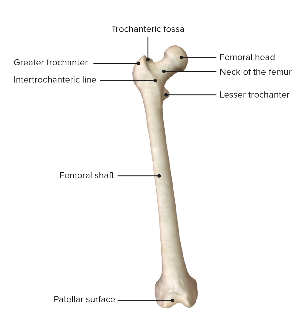
00:01 Now let's move on and look at the medial thigh muscles. 00:04 So the muscles that formed the medial compartment of the thigh. 00:08 Here we can see a whole range of muscles pectineus, obturator externus, adductor longus, adductor brevis, adductor Magnus, and gracilis, so a whole series of muscles here that are forming parts of the medial compartment of the thigh. 00:28 Let's have a look at pectineus. 00:30 Pectineus is emerging from the pectin pubis. 00:33 So this pubic line running across the pubic bone and it runs all the way down to the pectineal line of the femur. 00:42 So when we looked at the bony landmarks on the femur and we mentioned the pectineal line, that's because pectineus muscle inserts onto it. 00:49 Reminding ourselves this is the anterior surface of the right hip joint. 00:54 So pectineus is running from the pubic bone down to the femur. 00:58 It's associated with adduction. 01:01 So it helps to move the femur closer towards the midline and also flexion of the hip joint because it's lying anterior to that hip joint. 01:10 So it adducts and it flexes the thigh at the hip joint. 01:15 If we then look at obturator externus, we can see obturator externus here is coming from the external surface of the obturator membrane and some of the surrounding bones. 01:26 This muscle passes posterior to the neck of the femur, and we can see it's attaching to the trochanteric fossa. 01:34 So this muscle is passing posterior to the femur. 01:37 This gives an indication of its movement because if it was to contract, it would laterally rotate the thigh at the hip joint because it's running posterior to that hip joint. 01:48 If we were to then move on and look at gracilis this is a very slim and slender muscle that is positioned quite superficially, very close to the skin on the medial aspect of the thigh. 01:59 We can see gracilis is coming down from the inferior pubic ramus and it's passing towards the medial aspect of the tibial tuberosity. 02:07 It's running very superficially though. 02:10 It has a couple of important movements, it helps to abduct the site at the hip joint, and it also helps to flex the leg at the knee joint. 02:19 The next muscle is a very big muscle. 02:21 It's the primary abductor of the thigh at the hip joint. 02:25 Here we can see a adductor Magnus from the anterior view. 02:29 And then if we spin it around, we see a adductor Magnus from the posterior view as well. 02:34 Posteriorly, there's two bits to it. 02:37 We've got an abductor part, which we see radiating from the issue of the pelvis all the way down to the femur. 02:44 We then have a portion that runs more directly down towards the distal aspect of the femur. 02:50 And here we can see both an abductor and a hamstring part. 02:54 As these two parts separate towards the distal aspect of the femur, we find the abductor hiatus and this is an important channel known as the abductor canal that allows the femoral nerve and vein to pass from the anterior compartment of the thigh into the popliteal fossa. 03:11 Throughout its course though, there are various holes allowing for perforating branches to come and really supply the depth of the thigh. 03:19 So let's have a look at the origin and the insertion of adductor Magnus. 03:23 Let's look at the abductor part, that's coming from the ischial tuberosity. 03:27 And it's radiating towards the femur where we can see it attaching to the linear aspera. 03:32 So that's the abductor part. 03:33 If we were to look at the hamstring part, that comes much further down and goes to the adductor tubercle on the femur, and it's originating from the ischial tuberosity. 03:42 So a very large muscle that has both adduct to and hamstring parts and you can see its broad origins and insertions there. 03:50 If we were to then have a look at the innovation, the hamstring parts of adductor Magnus is why the tibial nerve, whereas the abductor portion is via the obturator nerve. 04:00 If we look at the function of the hamstring part and the abductor part of adductor Magnus, then its name does help us a little bit. 04:07 That hamstring part because it doesn't cross the knee joint, doesn't have anything to do with the leg, but it does cross the hip joint and it's an important extensor of the thigh at the hip joint. 04:17 The abductor portion is obviously important with adopting the thigh at the hip joint. 04:23 Now let's have a look at some other abductor muscles. 04:25 We have adductor longus and adductor brevis. 04:28 If we look at the origins and insertions of adductor longus, we can see adductor longus is originating from the body of the pubis. 04:36 It then runs inferolaterally towards the linear aspera on the femur. 04:41 This muscle is associated with adduction of the thigh at the hip joint like the other muscles in this compartment. 04:48 We then have adductor brevis. 04:50 This is a smaller version of adductor longus. 04:52 And this also is coming from the pubic bone. 04:55 It comes from the body and inferior ramus of the pubis, and again it runs laterally towards the proximal parts of the linear aspera. 05:04 It also has a function in helping to abduct the thigh at the hip joint. 05:10 Innovation of the medial compartment is done to various muscles, so we've got obturator externus, abductor brevis, adductor longus, adductor Magnus, it's adductor part, and gracilis and these all integrated right in the obturator nerve. 05:23 So the obturator nerve is the primary adductor nerve of the thigh. 05:27 Damage to the obturator nerve therefore in the pelvis can limit this movement. 05:32 Pectineus and the adductor Magnus muscles hamstring part, while the pectineus is supplied by the femoral nerve and the adductor Magnus is hamstring part is supplied by the sciatic nerve.
About the Lecture
The lecture Medial Compartment of the Thigh by James Pickering, PhD is from the course Anatomy of the Thigh.
Included Quiz Questions
What is NOT a muscle of the medial thigh?
- Sartorius
- Pectineus
- Adductor longus
- Obturator externus
- Adductor brevis
What is the insertion site of the obturator externus?
- Trochanteric fossa
- External surface of obturator membrane
- Internal surface of obturator membrane
- Inferior pubic ramus
- Tibial tuberosity
What is the function of the obturator externus?
- Lateral rotation of the thigh at the hip joint
- Medial rotation of the thigh at the hip joint
- Flexion of knee joint
- Extension of knee joint
- Extension of hip
Customer reviews
5,0 of 5 stars
| 5 Stars |
|
5 |
| 4 Stars |
|
0 |
| 3 Stars |
|
0 |
| 2 Stars |
|
0 |
| 1 Star |
|
0 |


