Playlist
Show Playlist
Hide Playlist
Lobar Atelectasis


-
Slides Atelectasis.pdf
-
Download Lecture Overview
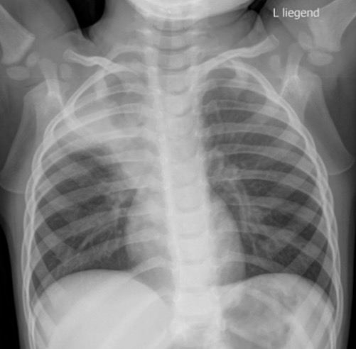
00:01 So let's take a look at some common patterns that you may encounter. 00:03 What do you see on this chest x-ray? So this is the finding here, this is the type of atelectasis. 00:11 So what kind of atelectasis do you think this is and where is it located? So this is right upper lobe collapse or right upper lobe atelectasis. 00:25 This results in a shift of the minor fissure superiorly. 00:28 So this is an example of the minor fissure where the area of consolidation stops and then you may have a shift of the trachea towards the side of collapse because it's a relatively small segment that trachea may or may not shift. 00:41 How does this picture differ from the one that you just saw? So you can see this pattern here, again it has an area of consolidation with very sharp margins that can be outlined by the screen line but it has a little bit of a bulge right here. 01:02 It almost looks like a backward S. This is the S sign of Golden and this is what a hilar mass resulting in right upper lobe collapse looks like so the hilar mass causes obstruction of the bronchus which then result in right upper lobe collapse. 01:17 So whenever you see an example of right upper lobe collapse, you wanna see if there's any kind of nodularity or mass surrounding the hilum which would represent the S sign of golden. 01:26 So let's take a look at this frontal and lateral view of the chest. 01:30 So here, we have an opacity with very sharp margins. 01:33 On the frontal view, it silhouettes the heart border and on the lateral view it seen overlapping the heart. 01:39 So this is an example of right middle lobe collapse. 01:43 Right middle lobe collapse typically silhouettes the right heart border. 01:46 It causes elevation of the hemidiaphragm and it causes a density on the lateral view that overlaps the heart. 01:53 So this is a very typical finding of right middle lobe collapse. 01:57 Let's take a look at this one here. 02:02 Again, we have a frontal and a lateral view and here is our abnormality. 02:08 So we have a density adjacent to the right heart border that seen posteriorly on the lateral view. So where could this be located? So this is an example of right lower lobe collapse. 02:25 So the pattern that you see is shifted the minor fissure down, so this is the minor fissure right here and it shifted downwards. 02:32 You have shift the heart towards the right or towards the side of collapse. 02:36 You have elevation of the right hemidiaphragm which we can see here, and you have silhouetting of the right hemidiaphragm. 02:44 You may see a density seen posterior to the heart on the lateral view. 02:49 So again to help you differentiate from a right middle lobe collapse. 02:54 On lateral view, the right middle lobe collapse will overlap the heart, while on a lateral view, the right lower lobe collapse will be seen posteriorly. 03:02 So let's take a look at this one, this one can be actually a little bit tricky. 03:08 So we have frontal and a lateral view of the chest. 03:11 So the entire left lung looks a little bit abnormal. 03:17 If you compare with the right it actually looks a little bit more hazy than the right lung does, and on the lateral view, you see a density anteriorly and you can see a fine line where the arrows are. 03:29 So this is an example of left upper lobe collapse. 03:33 This is actually a very easy to mis-finding so it's important to try to keep this one in mind. 03:38 You have a hazy density that surrounds the left hilum. 03:41 You may or may not the have elevation of the left hemidiaphragm. 03:44 You again may or may not have shift of the mediastinal structures to the left and the lateral view demonstrates a linear density that's posterior to the sternum. 03:53 Then you see essentially from top to bottom and this is a classic example of left upper lobe collapse. 03:58 So what do you see on this? This is another one that can be a little bit tricky. 04:04 So we only have a frontal view here and you see a density that's right behind the heart. 04:17 This is an example of left lower lobe collapse. 04:20 So the density is seen behind the heart which can make it hard to see. 04:23 You may have shift of the mediastinal fissure down and you have silhouetting of the left hemidiaphragm. 04:30 So again if you take a look here, we should be able to see the entire left hemidiaphragm. 04:34 However with the area of atelectasis part of it is silhouetting. 04:37 If you have a lateral view within the density will be posterior to the heart. 04:42 So how can we differentiate atelectasis and pneumonia? Both of them appear as an area of consolidation within the lung. 04:52 So atelectasis has very sharp borders as we saw on these examples while pneumonia has very hazy borders. 04:58 In atelectasis we would not see any air bronchograms while in pneumonia you do. 05:03 Atelectasis is volume loss while with pneumonia you really don't have any evidence of volume loss so you wouldn?t have shift of the mediastinal structures and you wouldn't have shift of the hemidiaphragm. 05:14 Both can produce the silhouette sign and both results in increase to density. 05:20 Atelectasis has a very rapid change over time while pneumonia has a slow change in time. 05:25 So if you're not sure which one you're looking at, if the patient has had recent prior images, it's helpful to compare to see if this came up quickly or if this was present for a long time. 05:35 So let's take a look at these two cases, which one is atelectasis and which one is pneumonia? We have the lateral view for both as well. 05:56 So the one on your left is the right middle lobe pneumonia because it has hazy margins, it's a hazy opacity and the one on the right is the right middle lobe atelectasis which has very sharp margins as you can see both appears to be dense on the radiograph and both silhouette the right heart border.
About the Lecture
The lecture Lobar Atelectasis by Hetal Verma, MD is from the course Thoracic Radiology. It contains the following chapters:
- Lobar Atelectasis
- Atelectasis vs. Pneumonia
Included Quiz Questions
What is the name of the radiographic sign highly suggestive of a hilar mass resulting in the collapse of the right upper lung lobe?
- S sign of golden
- Silhouette sign
- Loss of the more black sign
- Deep sulcus sign
- Spine sign
Which of the following is NOT a diagnostic feature of the right middle lobe collapse?
- Indistinct margins of the opacity
- Silhouetting of the right heart border on frontal view
- Elevation of the diaphragm
- Density overlying the heart on the lateral view
- Increased density on the radiograph
Chest X-ray reveals a density adjacent to the right heart border obscuring the medial right hemidiaphragm, inferior displacement of the right hilum, and shifting of the minor fissure down. What is the most likely diagnosis?
- Right lower lobe collapse
- Left upper lobe collapse
- Left lower lobe collapse
- Right lower lobe consolidation
- Right upper lobe collapse
Which feature differentiates atelectasis from pneumonia?
- Absence of air bronchograms
- Silhouette sign
- Increased density
- Shortness of breath
- Tachypnea
Author of lecture Lobar Atelectasis

Hetal Verma, MD
Customer reviews
5,0 of 5 stars
| 5 Stars |
|
1 |
| 4 Stars |
|
0 |
| 3 Stars |
|
0 |
| 2 Stars |
|
0 |
| 1 Star |
|
0 |
Dear Hetal Verma, Thanks for presenting good lecture I like it very much
