Playlist
Show Playlist
Hide Playlist
Ligaments and Menisci of the Knee
-
Slides Ligaments and Menisci of the Knee.pdf
-
Download Lecture Overview
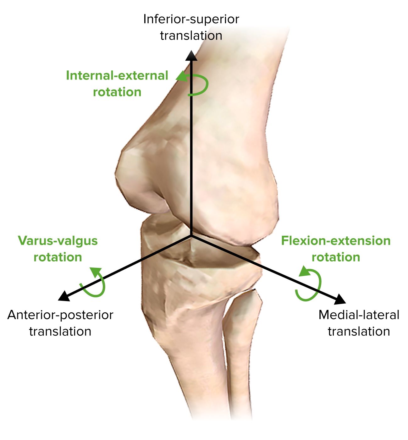
00:01 Now, let's look at the ligaments and the menisci of the knee joint. 00:06 So here we can need to add on a whole series of ligaments associated with the knee. 00:12 We'd have heard about these before when we talked about the muscles that act on the knee. 00:17 So here we have the Patellar ligament. 00:19 It's the continuation of quadriceps femoris tendon that's coming down from the anterior thigh. 00:24 It's running over the patella which forms within this tendon and then it passes down as the patellar ligament that attaches to the tibial tuberosity. 00:34 We can also see, if we look on this lateral aspect, here we can see the patellar ligament is anteriorly. 00:40 So this is the anterolateral view of a right knee, we can see quadriceps femoris superiorly. 00:47 But we can see running from the lateral condyle of the femur to the fibular, the fibular collateral ligaments or the lateral collateral ligament. 00:59 Hearing see it coming from the lateral femoral epicondyle. 01:02 And it's running down to the head of the fibular. 01:06 So here we see the fibular collateral ligament. 01:09 If we spin the knee around, we can see we have the equivalent on this medial side. 01:14 And this is the tibial collateral ligament. 01:17 Coming from the medial femoral epicondyle and attaching to the medial surface of the tibia, we can see the tibial collateral ligament. 01:26 These helped to stabilize the knee joint. 01:29 We also have running within that joint capsule, between the two condyles. 01:34 We have the anterior cruciate ligament, this is coming from the anterior part of the intercondylar area. 01:42 Remember we saw that when we looked at this tibial plateau previously. 01:47 So the anterior cruciate ligament is coming from the anterior part of the tibia and it passes posteriorly to attach to attach to the lateral wall of this intercondylar fossa. 01:59 Here we can see the posterior cruciate ligament. 02:02 And this ligament is going to run anteriorly forming a cross with its sibling, the anterior cruciate ligament. 02:10 So here we have the posterior cruciate ligament, emerging from the posterior part of the intercondylar area. 02:17 This then runs up to the medial wall of the intercondylar of fossa. 02:22 So the anterior cruciate ligament comes from the anterior aspect of the inter condyle area. 02:28 And the posterior cruciate ligament comes from the posterior part of the intercondylar area. 02:35 These ligaments are important in preventing the femur sliding off the surface of the tibial plateau. 02:41 So with forced movements, sometimes the femur can tend to slide off the tibial plateau. 02:47 These ligaments hold it in position to prevent the tibia and the femur from sliding against one another. 02:54 If we stay on this posterior surface, we can now see the oblique popliteal ligament. 03:00 This is passing down from the lateral condyle of the femur all the way to below the posterior margin of the head of the tibia. 03:09 So we can see that oblique popliteal ligament running in that direction. 03:13 It's also joined by a tendinous extension of that semimembranosus muscle. 03:18 And this again helps to reinforce that medial aspect of the knee, helping to stabilize it in position. 03:26 More on the lateral aspects, we have the arcuate popliteal ligament, and this is running from the head of the fibular all the way up to the lateral epicondyle of the femur. 03:36 These ligaments are very important both within the substance of the knee, and these ligaments peripherally on the lateral and medial margins in stabilizing the knee in position. 03:48 The knee is really important in your standing up posture, as it bears all the weight of both yourself, and that of gravity pushing down on your head and shoulders. 03:57 So it needs to maintain stability. 04:00 If we then look at the menisci, these are cartilaginous rings that sit on the tibial plateau. 04:06 So here again, we're looking at the anterior surface of the right knee, the femur has been elevated and we're left with the lateral meniscus. 04:15 On the opposite side, we have a medial meniscus. 04:18 Separating the two, we have that intercondylar area. 04:22 And most anteriorly we have that transverse ligament of the knee. 04:27 These are important in helping to increase bony congruency of this joint. 04:32 We had the we had the acetabulum labrum, which have similar function in the hip joint. 04:36 What these two do, the lateral and the medial menisci, is they surround some of the condyles of the femur. 04:44 Increasing the surface area and increase in the area for articulation between the tibial plateau and the femur. 04:52 Once again, in the standing upright position, they form these little cups for the femoral condyle to sit in and to maintain stability. 05:01 If we then look at the vascular supply of the knee joint, it's very much coming from the femoral artery, which gives rise to a series of branches as it descends. 05:10 It's not quite the popliteal branch, yet. 05:12 But here we've got an articular branch that's coming down from the femoral artery. 05:16 And we've also got a descending branch that's coming from the deep femoral artery. 05:21 These go on to help support an anastomotic ring around the knee joint. 05:26 As the femoral artery passes through the adductor canal, it becomes the popliteal artery, which gives rise to these four arteries. 05:34 These are your genicular arteries. 05:37 We have two superior and two inferior, that both come up from above or below the knee joint itself. 05:45 Above the knee joint, we have the superior lateral and superior medial genicular arteries. 05:51 And below the knee joint, we have the inferior lateral and the inferior medial genicular arteries. 05:58 We also have contributions once the popliteal artery is passed down through the popliteal fossa, it gives rise to the tibial artery. 06:06 And here we can see the anterior tibial recurrent artery. 06:10 We also, as its name suggests, will have a posterior tibial recurrent artery. 06:15 These supply lots of additional branches into this anastomotic loop, ensuring that the joint capsule around the knee receives the nutrients it needs to keep on working and remaining healthy. 06:29 Now let's have a look at the movements of the knee joint. 06:31 We can see here the tibia is moving posteriorly within the sagittal plane at the knee joint. 06:37 This is flexion of the tibia at the knee joint. 06:40 And then as it moves anteriorly within that sagittal plane, we can see it's extending back to this resting position. 06:48 Flexion and extension. 06:51 If we then look down onto the tibial plateau, superior view onto the right leg, we can actually appreciate here we have some lateral rotation, and then the opposite we'll have some medial rotation. 07:05 So external or internal, lateral or medial rotation of the tibia at the knee joint.
About the Lecture
The lecture Ligaments and Menisci of the Knee by James Pickering, PhD is from the course Joints of the Lower Limbs.
Included Quiz Questions
What is one attachment site for the fibular collateral ligament?
- Lateral femoral epicondyle
- Medial femoral epicondyle
- Medial surface of the tibia
- Medial surface of the fibula
- Patella
What is one attachment site of the anterior cruciate ligament?
- Lateral wall of intercondylar fossa
- Posterior part of intercondylar area
- Medial wall of intercondylar fossa
- Medial part of intercondylar area
- Head of the fibula
What ligament is closely associated with the tendinous extension of the semimembranosus muscle?
- Oblique popliteal
- Anterior cruciate
- Posterior cruciate
- Lateral collateral
- Medial collateral
Which artery does NOT supply blood to the knee?
- Posterior lateral genicular
- Superior lateral genicular
- Superior medial genicular
- Inferior lateral genicular
- Inferior medial genicular
Customer reviews
5,0 of 5 stars
| 5 Stars |
|
5 |
| 4 Stars |
|
0 |
| 3 Stars |
|
0 |
| 2 Stars |
|
0 |
| 1 Star |
|
0 |


