Playlist
Show Playlist
Hide Playlist
Knee Joint

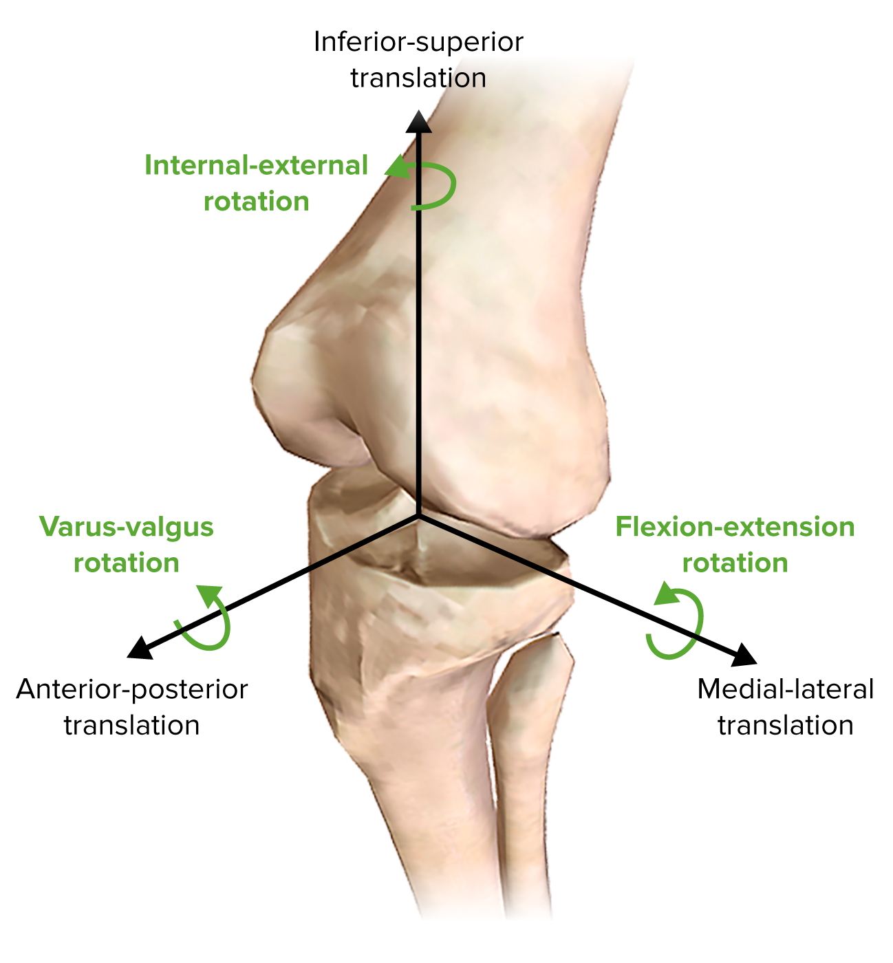

00:01 Now let's have a look at the knee joint. 00:04 The knee joint is a hinge Synovial joint. 00:07 And here we're looking at the medial surface of the right knee. 00:12 We can see the patella is positioned anteriorly. 00:15 Here we can see some articular surfaces of the knee joint and they occur between the tibia, the femur and the patella. 00:23 So here we have the femur, we have it's dilated condyles - the medial and lateral femoral condyles. 00:30 And these are going to articulate with the medial and lateral tibial condyles on the tibia. 00:36 These are the femoral tibial articulations. 00:40 We also, on the anterior surface of the femur, have the patella surface. 00:44 And this interacts or articulates with the posterior surface of the patella. 00:50 This is the Femoropatellar articulation. 00:54 Surrounding the knee joint, we find a joint capsule. 00:58 And this is quite an extensive joint capsule. 01:01 A fibrous membrane that's quite loose to allow great flexibility of the knee. 01:05 And that surrounds both the condyles of the femur and the tibia. 01:11 The superior attachment of this joint capsule are the margins of the articular surfaces of the condyles. 01:17 And then inferiorly, it's similarly the margins of the tibial articular surfaces. 01:23 Lining the inside of this joint capsule is an important Synovial membrane. 01:28 And that enables the bony surfaces to glide smoothly against one another. 01:32 Its margins are very similar to that of the fibrous joint capsule. 01:36 We have margins around the articular surfaces. 01:39 And it also forms attachments on the outer margins of the menisci. 01:43 If we rotate and look at the knee posteriorly, we can see we have the synovial membranes attached here, and also in between the two condyles. 01:53 But outside of the synovial membranes, do we find the cruciate ligaments, we'll talk about those later on. 02:00 We can then see some specific Alar folds and an Infrapatellar fold formed by the synovial membranes around the posterior aspect of the knee. 02:10 If we then look, we can see there's various extensions of this Synovial membrane that form various Bursa. 02:17 Here we have the Suprapatellar bursa. 02:19 Here we have some tendon of popliteus muscle running over a Subpopliteal recess. 02:26 Now, what these do is these create little pouches of synovial fluid lined by this Synovial membrane, and they help to prevent friction of tendons that run very closely and alongside various bony structures. 02:40 So these are important as they help tendons to run smoothly over their bony surfaces.
About the Lecture
The lecture Knee Joint by James Pickering, PhD is from the course Joints of the Lower Limbs.
Included Quiz Questions
What is the location of the cruciate ligaments in relation to the synovial membrane?
- The cruciate ligaments are outside the synovial membrane.
- The cruciate ligaments are within the synovial membrane.
- The cruciate ligaments are lateral to the synovial membrane.
- The cruciate ligaments are inferior to the synovial membrane.
- The cruciate ligaments are medial to the synovial membrane.
What are extensions of the synovial membrane? Select all that apply.
- Suprapatellar bursa
- Subpopliteal recess
- Popliteus bursa
- Patellar bursa
- Patellar recess
Author of lecture Knee Joint

James Pickering, PhD
Customer reviews
5,0 of 5 stars
| 5 Stars |
|
5 |
| 4 Stars |
|
0 |
| 3 Stars |
|
0 |
| 2 Stars |
|
0 |
| 1 Star |
|
0 |
