Playlist
Show Playlist
Hide Playlist
Joints of the Hand

-
Slide Joints of the Hand.pdf
-
Download Lecture Overview
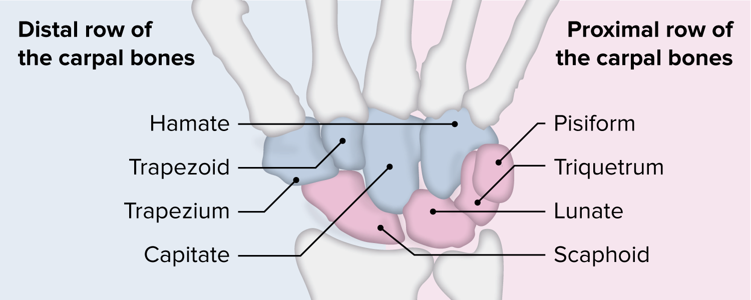
00:01 So, now, let's move most distally away from the shoulder joints, the glenohumeral joints and the elbow joint. 00:08 And look at the numerous joints associated with the hand. 00:11 And there's a lot here. Let's first of all start by looking at the wrist joint. 00:15 And here on the anterior surface of the wrist on the right-hand side, we see the radius. 00:19 The left-hand side, we see the ulna. 00:22 We can see the articulation of the wrist joint here via the carpal bones. 00:26 Here, we can then see the carpometacarpal joints where the carpal bones are connected to the metacarpals. 00:33 And then, most distally, we'll see the interphalangeal joints. 00:37 We'll come to those in a moment. So, let's have a look at the articular surfaces. 00:42 Here, we've got the articular surfaces at the distal end of the radius and the ulna. 00:46 We can see the articular discs. And these are articulating with the scaphoid and the lunate bones for the radius and the triquetrum for the ulna. 00:56 So, we can see, we have a whole series of these carpal bones that are sitting against the distal end of the radius and the ulnar. And they form these articulations. 01:07 There's a number of numerous ligaments associated to these bones and that really is to hold these bones in place. If you think we've got eight pebbles really connecting the distal end of the radius and the ulna to the carpal bones in the hand. 01:24 Obviously, we have these to enable a wide range of movement and flexibility within our wrist joints. 01:30 But that means we have to have a number of ligaments to hold these in place. 01:34 So, here, we can see a whole series of radiocarpal ligaments. 01:38 Positioned on the palmar surface so that the palmar radiocarpal ligaments. 01:43 We have some specific ones here. We can see the radial collateral ligament and then, we have some equivalent on the ulna side, these palmar ulnocarpal ligaments, helping to hold the carpal bones to the ulna. 01:56 And again, we have an ulnar collateral ligament observed as well. 02:00 These ligaments really are here to hold those bones together and hold them against the articular surfaces of the radius and the ulna. 02:08 If you were to look on the posterior aspect, we can again pick out the radial and ulnar collateral ligaments. 02:14 But importantly, we can see this quite prominent ligament which is the dorsal radiocarpal ligaments. 02:20 We can see these again running similar to the palmar ones but connecting the radius to these carpal bones. 02:27 These are really important because they allow a wide range of movement. 02:30 So, here, we have abduction and here, we have adduction. 02:34 Similarly, we can see we have flexion and extension. 02:38 So, a wide range of movements permitted by these interconnected bony structures between the radius, ulna, and the carpal bones, supported by those numerous ligaments. 02:51 If we then look at the joints between the carpal bones themselves, we have what are known as intercarpal joints. 02:57 Again, these are really just supported by a number of ligaments which are primarily concerned with holding these bones together. 03:04 Holding them together so their articulations can take place. 03:09 So, now, let's concentrate on the carpometacarpal joints. 03:12 These are associated with the carpal bones proximally and the metacarpals distally. 03:19 So, here, we can see we have that distal row of carpal bones. 03:23 We can see these four bones situated here and they're going to articulate with the proximal end of those five metacarpals. We can see them here, one, two, three, four five. 03:35 These are the five metacarpals. We have quite limited movement in this space but we have wide range of mobility within the first one. 03:44 That means the thumb can assume a lot more different positions compared to the digits two, three, four, and five. 03:51 And it's important to appreciate these differences in movement. 03:55 So, now, let's look at the movement of the carpometacarpal joints and pay particular attention to the first digit, the thumb, first of all. 04:04 Now, you have to remember, if you were standing in the anatomical position, then, flexion and extension really goes on through the sagittal plain. 04:11 So, flexion and extension of your fingers, flexion and extension of your forearm at the elbow joint, these are occurring in the sagittal plain whereas abduction and adduction of your fingers or abduction and adduction of your arm, is occurring in the coronal plane. 04:30 We have to remember for the thumb, we have to realize that it's really being rotated 90 degrees. 04:37 So, if you think of the orientation of your fingers and the thumb, you can see the thumb appears to have been rotated 90 degrees. 04:44 So, where flexion and extension of your fingers occurs in that sagittal plane, if we imagine we've rotated that plane because of the thumb, extension and flexion and extension of the thumb occurs in the coronal plane. 05:00 So, as we can see in the image here, flexion of the thumb moves across in the coronal plane across the surface of the palm. 05:08 Extension then moves again in the coronal plane but away from the palm because the planes in which it moves has been rotated. 05:17 That means that again, in the resting position, if we were to abduct and adduct our thumb, we would now move abduction and adduction as if we were in the sagittal plane because it's being rotated. 05:31 So, for the rest of the body, abduction, adduction occurs in the coronal plane. 05:35 Now, for the thumb, it works in the sagittal plane. 05:39 So, now, we have abduction which moves it away and adduction which brings it towards the side of the palm. 05:47 So, abduction and adduction is now occurring in the sagittal plane whereas flexion and extension works in the coronal plane. 05:56 Because if you look at the thumb, it's clearly been rotated 90 degrees. 06:01 So, these movements in the muscles that move the thumb in this direction is important to recognize how different they are. 06:08 Also occurring within the carpometacarpal joints is we have rotation and circumduction, allowing the wrist to move.
About the Lecture
The lecture Joints of the Hand by James Pickering, PhD is from the course Joints of the Upper Limbs.
Included Quiz Questions
Which statements concerning the scaphoid bone are correct? Select all that apply.
- It can be palpated in the anatomical snuff box.
- It articulates with the ulna.
- It is boat-shaped.
- It articulates with the trapezium.
- It is prone to fracture in a fall on the outstretched hand.
Which bones take part in articulation in the wrist joint? Select all that apply.
- Scaphoid
- Pisiform
- Lunate
- Triquetrum
- Radius
The ulnar collateral ligament of the wrist runs from the ulnar styloid process to which bone?
- Triquetrum
- Scaphoid
- Lunate
- Capitate
- Hamate
In which plane do thumb adduction and abduction occur?
- Sagittal
- Coronal
- Axial
- Transverse
Author of lecture Joints of the Hand

James Pickering, PhD
Customer reviews
5,0 of 5 stars
| 5 Stars |
|
5 |
| 4 Stars |
|
0 |
| 3 Stars |
|
0 |
| 2 Stars |
|
0 |
| 1 Star |
|
0 |
