Playlist
Show Playlist
Hide Playlist
Joints of the Foot

-
Slide Joints of the Foot.pdf
-
Download Lecture Overview
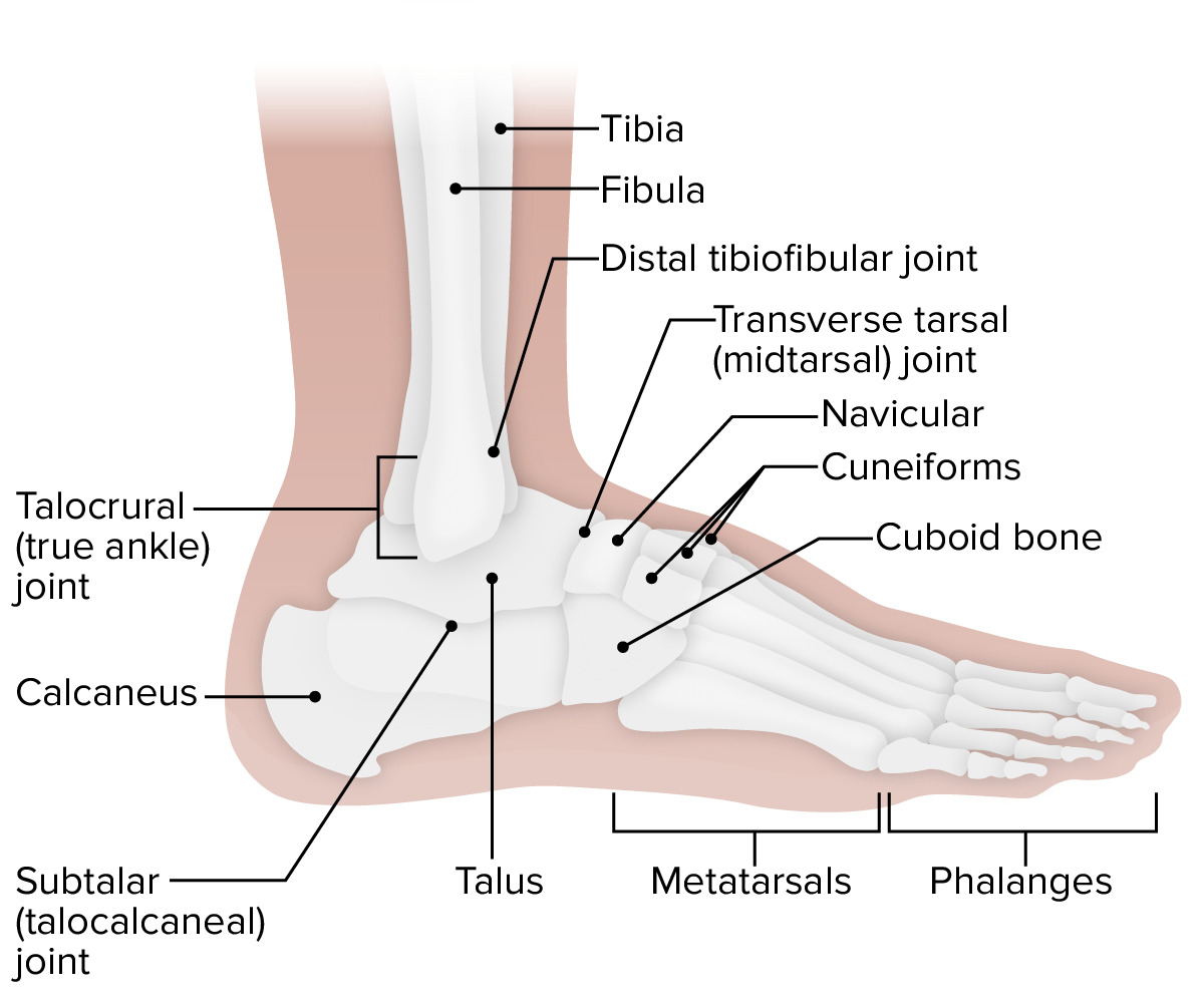
00:01 So now let's have a look at some joints within the foot. 00:05 So here we're going to look at some subtalar joints. 00:09 And these joints really exist between a number of bones that sit inferior to the tibia and fibular within the tarsal bones of the foot. 00:18 And these really form plane synovial joints. 00:21 And we can see a whole series of these occurring. 00:24 Let's have a look at the subtalar joint, first of all. 00:27 It's running from the inferior surface of the talus, and it's articulating with the superior surface of the calcaneus. 00:34 So here we have the subtalar joint, it's inferior to the talus. 00:39 The ligaments are there to reinforce this joint and help to hold it in position. 00:43 Here we have the interosseous talocalcaneal ligament. 00:46 And here we have the lateral talocalcaneal ligament, helping to hold the talus against the calcaneal. 00:54 These happen more anteriorly. 00:56 Posteriorly, we have the posterior talocalcaneal ligaments, and then right on the medial side, we'll have the medial talocalcaneal ligament. 01:05 So four talocalcaneal ligaments helping to hold the talus and the calcaneus bones together. 01:12 Now we're gonna have a look at a series of joints that really are associated with the tarsal bones moving anteriorly or more distally towards the metatarsals. 01:21 There's quite a lot here. 01:23 And the words can seem slightly confusing, but really, the words are just indicating the joints in which the bones are articulating. 01:31 So the talocalcaneonavicular joint is going to exist between the talus, the calcaneus, and the navicular bone, the talocalcaneonavicular joint. 01:43 Here we can see the articular surfaces. 01:45 Here we see the head of the talus articulating with that articular surface of the navicular bone. 01:51 And here we can see an articular surface of the calcaneus. 01:54 And those three are going to come together to form the talocalcaneonavicular joint. 02:00 Again, we'll have some ligaments that help to reinforce this. 02:03 Again, we have the interosseous talocalcaneal ligament. 02:06 And now we have the bifurcates ligament. 02:10 This is the calcaneonavicular and the calcaneocuboid. 02:14 Two ligaments, essentially running from the calcaneus to the navicular, the calcaneus to the cuboid bone, the bifurcate ligament. 02:23 Running more dorsally, we have the dorsal talonavicular ligament, we can see here. 02:28 And here we see a spring type ligament. 02:31 And this is really important in giving some mobility and stretch, especially important as we're walking and running to maintain momentum. 02:39 And this is the plantar talonavicular ligament. 02:42 It's the plantar as it helps to plant the foot and it's the talonavicular passing between the talus and then the navicular bones. 02:50 Now let's have a look more laterally. 02:52 And we have the calcaneocuboid joint. 02:55 This is a synovial saddle joint that's articulating between the articular surface of the calcaneus with the posterior articular surface of the cuboid bone. 03:05 So here we can see the calcaneocuboid joint. 03:08 The ligament that's associated with this is again part of that bifurcated ligament, that bifurcated ligaments. 03:16 And that's the calcaneocuboid ligament, we can see here. 03:19 Running between the calcaneus and the cuboid bones. 03:23 We also running underneath this, have a dorsal calcaneocuboid ligament. 03:28 And that's running on the dorsal aspect of this joint. 03:32 If we have a dorsal one, the likelihood is we'll have a plantar to one and here we can see that plantar calcaneocuboid ligamen. 03:39 So two ligaments there which are again helping to hold these bones together. 03:44 Let's have a look at some classification now of these joints. 03:48 We've spoken a lot about all of the joints and the various bones they're running up against. 03:53 But actually what's the sort of function we have here? When we look at the functional subtalar joints, that is the talocalcaneal parts of the talocalcaneonavicular joint and the subtalar joint. 04:07 These are functional in that is there's a certain degree of movement that can occur at these joints to aid with mobility, to aid also with pressure compression, to give some spring when we're landing, and walking on our feet. 04:20 Then we have these transverse tarsal joints moving more anteriorly or moving more distally. 04:26 We have the talonavicular part of the talocalcaneonavicular joint. 04:32 And we also have the calcaneocuboid joints. 04:35 And these are running transversely across the tarsals of the foot. 04:41 Movement of the foot joints is relatively minor. 04:44 We just have inversion, and we have eversion. 04:47 This is when you lift either your big toe, inversion. 04:50 Or lift your little toe, eversion, off the ground. 04:53 And this happens when you're walking naturally. 04:57 There's a minor movement that can occur within the foot and that's not that substantial, but it's abduction and adduction. 05:03 And that is moving the foot both medially and laterally. 05:07 Again, very small, minimal movements of the foot but they're important in helping your foot assume different positions as you're walking around. 05:14 Some primary movements that you'll be familiar with is here we have dorsiflexion and plantarflexion. 05:20 And these help to spring us out forward as we're walking or running. 05:25 Just for completion, there's a few other intertarsal joints, but these don't have much function, really, they just holding the feet together, holding the foot bones together. 05:35 And this is the naviculocuneiform joint, the cuboidnavicular joint, the cuneocuboid joints, and the intercuneiform joints. 05:46 So those more distantly positioned tarsal bones. 05:49 They have also joined together, but you don't actually have that much movement that occurs between them. 05:55 We then to move even further distally and we find we have joints of the toes. 05:59 These are very similar to the joints we had of the digits in the hand. 06:03 So we have joints between the metatarsals and the proximal phalanges. 06:07 These are the metatarophalangeal joints, and we also have the interphalangeal joints that occur between the various phalanges. 06:15 One on the first digit, the big toe, and two for each of the remaining digits, two through two, five.
About the Lecture
The lecture Joints of the Foot by James Pickering, PhD is from the course Joints of the Lower Limbs.
Included Quiz Questions
How is the subtalar joint best classified?
- Plane synovial joint
- Syndesmosis joint
- Ball-and-socket joint
- Hinge joint
- Fibrous joint
How many ligaments support the talus and calcaneus?
- 4
- 5
- 6
- 2
- 3
What compromises the bifurcate ligament? Select all that apply.
- Calcaneonavicular
- Calcaneocuboid
- Dorsal talonavicular
- Plantar talonavicular
- Spring
What are the "functional" subtalar joints? Select all that apply.
- Talocalcaneal part of the talocalcaneonavicular joint
- Subtalar joint
- Calcaneocuboid joint
- Talonavicular part of the talocalcaneonavicular joint
Author of lecture Joints of the Foot

James Pickering, PhD
Customer reviews
5,0 of 5 stars
| 5 Stars |
|
5 |
| 4 Stars |
|
0 |
| 3 Stars |
|
0 |
| 2 Stars |
|
0 |
| 1 Star |
|
0 |
