Playlist
Show Playlist
Hide Playlist
Joints and Ligaments of the Back

-
Slides Anatomy Joints Ligaments of the Back.pdf
-
Download Lecture Overview
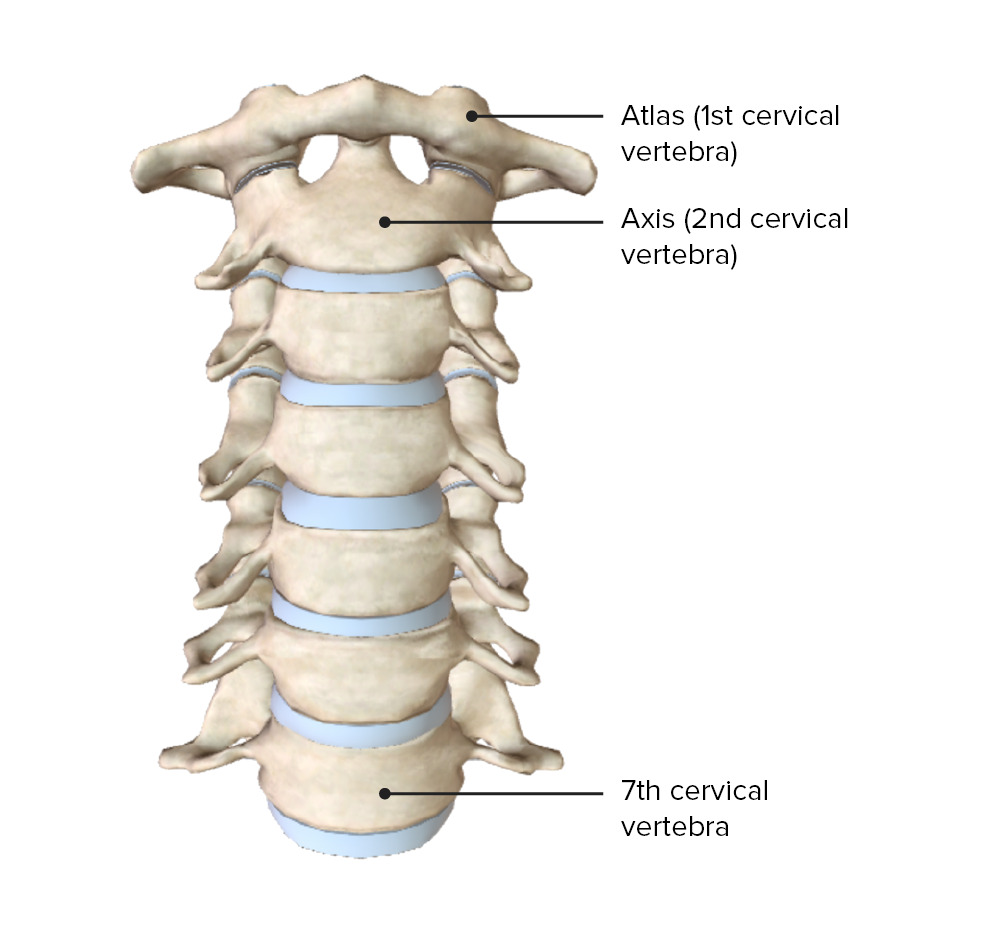
00:01 Now we're going to take a look at the joints and ligaments of the back that allow the back to move in so many different combinations of movements. 00:11 We'll start with a lateral view here of an intervertebral disc connecting the adjacent vertebral bodies. 00:20 This is a special type of joint called a symphysis. 00:23 And it's not the highly movable joint that you expect in what we call synovial joint, such as the elbow or the knees. 00:32 But it also provides a lot of support and a lot of shock absorption. 00:40 If we look more posterior in the vertebral arch, we see the inferior articular facet of one vertebra interacting with the superior articular facet of the vertebra below. 00:52 And together these facets form the facet joint or is like apophyseal joints. And these are synovial joints. 01:04 Synovial joints are joints that have a capsule and a cavity filled with joint fluid. 01:12 We call that the synovial cavity in the synovial fluid. 01:16 And these synovial joints are the ones that have a greater degree of movement. 01:22 So here we see these joints together in action. 01:26 We have flexion, which is really bending forward and extension just really bending backward. 01:32 And we have lateral flexion in either direction. 01:35 And they're fairly limited in their movement. 01:39 That's because we have the spinal cord. 01:41 We don't want to bend too far in any one direction, because then we cause damage to the spinal cord or its nerves. 01:49 So that brings us to ligaments to help provide structure and strength and stability. 01:55 So if we look at the base of the skull, going all the way down to the anterior surface of the sacrum inferiorly. 02:04 All of the anterior surface of the vertebral bodies and the intervertebral discs are covered by the anterior longitudinal ligament. 02:14 So it's a very descriptive term tells us where it is in which direction it's running. 02:19 Similarly, there's a structure up in the top of the cervical vertebra called the tectorial membrane. 02:27 And all along the posterior surfaces of the intervertebral discs and bodies run the posterior longitudinal ligament. 02:35 Really just the equivalent of the anterior longitudinal ligament on the other side of the vertebral bodies. 02:42 If we zoom in from a posterior point of view, and we look at adjacent laminae, which again, are the structures that connect the spinous process to the rest of vertebral arches. 02:54 We have these ligaments called ligamentum flavum. 02:57 It's a funny name and it's actually shown here in yellow because flavum means yellow, and it gets this yellowish appearance because of the presence of elastin fibers. 03:07 We swim around to a lateral view. 03:10 We see adjacent spinous processes connected by interspinous ligaments. 03:17 And if we look very, very posterior to the posterior most tips of the spinous processes, we see a ligament running along those tips called the supraspinous ligament. 03:30 As we move superiorly towards the cervical region, we see the spinous processes of the lower cervical vertebra here, going all the way up to the base of the skull. 03:42 And there's this free edge of ligament forming something about triangular shape. 03:46 And we call this the ligament of nuchae a referring to neck and as you might guess as you follow it down it becomes continuous with the supraspinous ligament.
About the Lecture
The lecture Joints and Ligaments of the Back by Darren Salmi, MD, MS is from the course Back Anatomy.
Included Quiz Questions
What joint is formed by the inferior and superior articular facets?
- Zygapophyseal joint
- Saddle joint
- Anterior joint
- Condyloid joint
- Pivot joint
What causes the ligamentum flavum to be yellowish in color?
- Presence of elastin
- Presence of collagen
- Presence of condyloid
- High-fiber diet
- High-protein diet
Author of lecture Joints and Ligaments of the Back

Darren Salmi, MD, MS
Customer reviews
5,0 of 5 stars
| 5 Stars |
|
5 |
| 4 Stars |
|
0 |
| 3 Stars |
|
0 |
| 2 Stars |
|
0 |
| 1 Star |
|
0 |
