Playlist
Show Playlist
Hide Playlist
Ischioanal Fossa

-
Slides Ischioanal Fossa.pdf
-
Download Lecture Overview
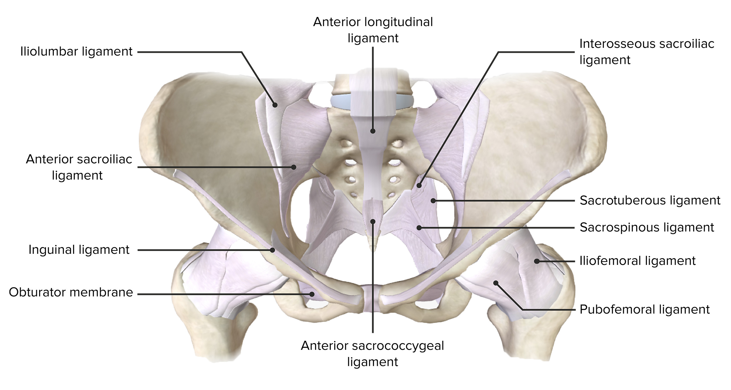
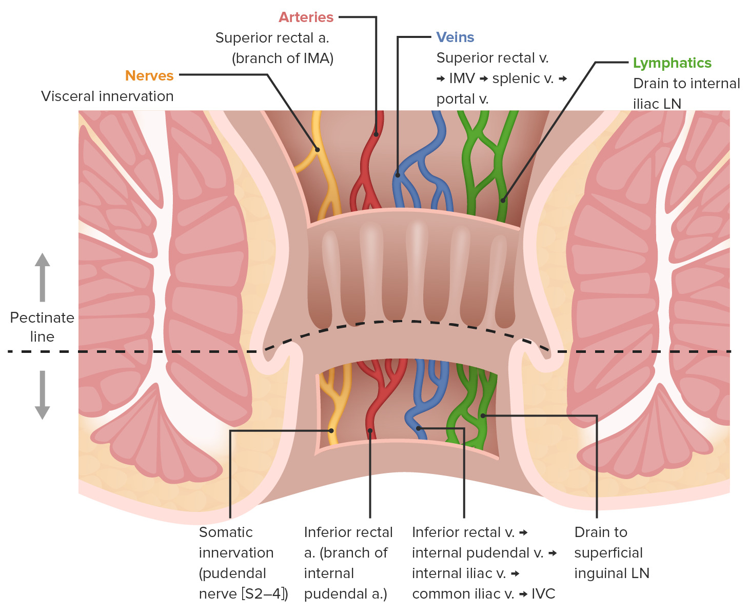
00:01 Now, let's have a look at the ischio-anal fossa in more detail. 00:05 So, here we're looking at the perineum from the posterior aspect. 00:09 We can see the sacrotuberous ligament is here. 00:12 We can see it's been removed. 00:14 So, there's the position of the sacred tuberous ligament. 00:16 You see also here we have obturator internus. 00:20 Remember, obturator internus is on the medial aspect of the obturator foramen, which we can see here. 00:27 So, let's just remove that again. 00:29 Here we've got obturator internus muscle sitting against the obturator membrane within the obturator foramen. 00:35 It's giving its long tendon out towards the greater trochanter of the femur. 00:40 And here we can see obturator internus. 00:42 So, this is an important landmark we'll come back to in a moment. 00:45 We can also see we've got the bone of the ischium highlighted here in green. 00:50 And these three structures from the lateral wall of the ischioanal fossa. 00:56 So, if you remember, the in the midline, we can see the bottom of the screen we have the external anal sphincter. 01:02 So, that's going to be in the midline. 01:04 So, the lateral wall is now formed by those three structures. 01:08 We've got the ischium, we've got obturator internus, and then we've got the sacred tuberous ligament. 01:13 These forming the lateral wall of the ischio-anal fossa. 01:18 Levator ani we can see there that is going to form alongside the external anal sphincter that's going to form the medial wall. 01:25 So, what we have is a relatively straight lateral wall, and then a angled medial wall. 01:33 And that creates this wedge shaped ischio-anal fossa, either side of the anus. 01:39 This wedge shaped structure is going to be filled with fat, which allows for expansion during defecation. 01:46 Expansion of the anal canal. 01:49 So, if we have a closer look at the ischio-anal fossa we can start looking at some various spaces. 01:56 Here we have the anterior recess of the issue of anal fossa. 02:00 Here we have the lateral wall of obturator internus. 02:03 Here we have the medial wall with levator ani muscles. 02:06 And here we can see the floor being the deep perineal pouch. 02:11 So this ischio-anal fossa is an important space. 02:15 We have two of them that sit either side of the anus, and they're mostly fat filled. 02:20 Now, let's look in more detail at the lateral wall of the ischio-anal fossa. 02:26 So, once again, let's orientate ourselves. 02:28 We're looking at the posterior aspects of the pelvis here. 02:32 So we can see the sacrum and coccyx quite clearly. 02:35 We can see the sacred tuberous ligament passing down from the sacrum to the ischial tuberosity. 02:41 And we can also now importantly, add in the sacrospinous ligament. 02:46 So, the sacrospinous ligament, remember is going from the sacrum to the ischial spine. 02:51 So sacrotuberous ligament here, sacrum to the ischial tuberosity. 02:55 And here's the sacrospinous ligament, sacrum to the ischial spine. 03:00 Here once again, we can bring in the space, which is the ischioanal fossa. 03:06 So what we're trying to describe here is how neurovascular structures that are emerging into the pelvis, but a destined for the perineum can actually make their way to the perineum without penetrating the pelvic floor. 03:21 So, the pelvic floor closes off the perineum from the pelvis. 03:26 So, the only way that structures can pass into the perineum is if they leave the pelvis. 03:33 They leave the pelvis to then go around the edge of the pelvic floor muscles. 03:38 So, then enter superficial to the pelvic floor muscles into that space, which is the perineum. 03:44 And in this case, how they're passing from the pelvis into the ischioanal fossa, that most posterior aspect of the perineum. 03:54 So, previously, we spoken about the various parameter within the pelvis. 03:58 And here we've highlighted the greater sciatic foramen. 04:02 And passing out of the greater sciatic foramen are some important neurovascular structures. 04:07 Here we can see the internal pudendal artery and vein, and the pudendal nerve. 04:13 What happens if I just bring those up again slowly, is for example, the internal pudendal artery, which is going to be emerging from branches of the internal iliac artery is going to pass out of the pelvis through the greater sciatic foramen. 04:28 As it passes out of the pelvis and the greater sciatic foramen, it loops around the sacrospinous ligament. 04:36 And as it loops around the sacrospinous ligament, it can now run a superficial to the pelvic floor muscles. 04:45 As it's running superficial to the pelvic floor muscles, it finds itself within the perineum. 04:52 So, the main blood supply to the perineum, the main venous drainage and the main nerve supply is the internal pudendal artery, the internal pudendal vein and the pudendal nerve. 05:03 And here we can see how they've left the pelvis via the greater sciatic foramen, Then they've cost around the sacrospinous ligament to now enter into the perineum. 05:13 They've gone around the free edge of the pelvic floor muscles, allowing them then to enter into the perineum. 05:21 What they've done is when they pass towards the ischioanal fossa is they give rise to branches that go and supply structures within it. 05:28 And in this case, one of the first structures they pass by is the external anal sphincter. 05:35 So here we can see coming from this neurovascular bundle, we have the inferior rectal vein, artery, and nerve. 05:42 As the structures run alongside the ischiopubic ramus and give off the inferior rectal vein, artery, and nerve. 05:49 They're running in a very important canal. 05:52 And this is known as the pudendal canal. 05:54 In some textbooks, you know as the alcock canal. 05:57 But its commonly call it the pudendal canal. 06:00 A little passageway that allows those three neurovascular structures to pass alongside the ischiopubic ramus. 06:06 Once they've passed alongside the ischiopubic ramus within the pudendal canal, they've given off these branches that run through the fat field ischio-anal fossa.
About the Lecture
The lecture Ischioanal Fossa by James Pickering, PhD is from the course Perineum.
Included Quiz Questions
What forms the lateral wall of the ischioanal fossa? Select all that apply.
- Sacrotuberous ligament
- Obturator internus
- Ischium
- External anal sphincter
- Levator ani
What forms the medial wall of the ischioanal fossa? Select all that apply.
- Levator ani
- External anal sphincter
- Sacrotuberous ligament
- Obturator internus
- Obturator externus
Author of lecture Ischioanal Fossa

James Pickering, PhD
Customer reviews
5,0 of 5 stars
| 5 Stars |
|
5 |
| 4 Stars |
|
0 |
| 3 Stars |
|
0 |
| 2 Stars |
|
0 |
| 1 Star |
|
0 |
