Playlist
Show Playlist
Hide Playlist
Inguinal Canal: Overview

-
Slides Inguinal Canal Overview.pdf
-
Download Lecture Overview
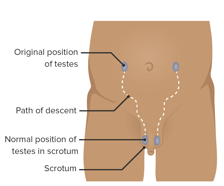
00:01 So let's now have a look at the formation and some of the contents of the inguinal canal. 00:06 It is a particularly complex layer, and you will need to understand the muscles of the anterolateral abdominal wall because I will be referring to them in great detail. 00:16 But here we can see a slightly close up cartoon version of the inguinal canal, the inguinal region. 00:23 Most superficially we can see in green, we have a layer of anterolateral abdominal wall. 00:29 And that is the external oblique muscle. 00:33 So here we can see external oblique muscle. 00:37 Deep to external oblique muscle you won't be surprised to see we have internal oblique muscle. And you can see that here. 00:44 The third muscle which was spoken about previously, is transverse abdominis. 00:49 So we have those three important muscle layers that you'll be familiar with from previous topics. 00:55 We have external oblique muscle most superficially, then deeper, internal oblique; and then deeper steel, we have transversus abdominis. 01:05 Finally, we have transversalis fascia, and then we'll have a layer of parietal peritoneum. 01:11 So these are important layers of the anterolateral abdominal wall. 01:15 And they're important in forming the inguinal canal in various different ways. 01:22 Previously, I mentioned really the entrance to the inguinal canal, if you're imagining we're passing from inside to out then we can call this the entrance. 01:30 Here we have the deep inguinal ring. 01:33 And the deep inguinal ring we can see is positioned really as an opening around transversalis fascia. 01:40 We'll come to its formation the moment or two. 01:42 But it's opening really within transversalis fascia there, and that's the deep inguinal ring. 01:48 We can see the canal then passes through. 01:52 And what actually happens, and we have to talk about embryology here. 01:55 Is if you imagine you have a structure. 01:57 Let's think of the testes and the biological male. 01:59 It's originating on the posterior abdominal wall, and it's destined in grown biologically male adults to be external. 02:08 So it needs to pass out through the abdomen. 02:12 And as it's passing out through the abdomen, it's taking with it various layers of the abdominal wall. 02:19 So what happens is the testes is pushing through the parietal peritoneum and pushing through transversalis fascia. 02:26 So as we can see here, that layer is coming through as the testee passes through this wall, and it's taking with it that layer of transversalis fascia tissue. 02:38 Here, we can now see that it's running underneath transversalis transversus abdominis muscle. 02:45 So remember, transversus abdominis muscle ran transversely across to the midline in the mid linea alba. 02:52 Now we can see the testes has taking its course, underneath transversus abdominis. 02:58 We can see the tendon of transverse abdominis actually carries on. 03:03 And now because we're below the level of the arcuate line, we can see its tendon runs anterior to rectus abdominis muscle. 03:12 So now we can see transversus abdominis is running towards the midline, anterior to rectus abdominis and it forms a region known as the conjoint tendon. 03:22 We'll come back to the conjoint tendon in a moment. 03:25 But you can see now clearly that the conjoint tendon is formed by both transversus abdominis and also internal oblique aponeurosis. 03:34 You can then see how the inguinal canal carries on and it's passing through internal oblique muscle. 03:42 Here we can see internal oblique muscle. 03:44 Now, this is important. Because the spermatic cord, which is what we're talking really here, as a large structure passing through the inguinal ligament. 03:54 It's no less significant than the female, the biological female. 03:58 But because the structure is much less inside, you only have the round ligament in the female. 04:05 In the male, you have the testes passing through. 04:08 So here we can see the testes now has taken a layer of transversalis fascia as it's passed through. 04:15 It hasn't taken a layer of transversus abdominis because it's passed underneath that muscle. 04:23 So that is now forming what we could call the roof of the canal. 04:27 But it's passed through internal oblique, so it's penetrated transversalis fascia, it's no penetrated internal oblique. 04:36 And internal oblique is not a layer of fascia like transversalis fascia is. 04:40 So as it's passed through this layer of muscle of internal oblique, it takes with it some muscle fibers, and this is the cremaster muscle. 04:49 So now that testes that's passed through, and in its wake, you've got various blood vessels and nerves and the vas deferens, those are all now covered by transversalis fascia and they're covered by the cremaster muscle. 05:03 We can then see the testes then penetrates again not the muscle but the aponeurosis of external oblique and that passing through of external oblique forms a superficial inguinal ring. Here we can see external oblique. 05:18 So here if we just have a closer look at the superficial inguinal ring. 05:23 We can see we have the superficial inguinal ring formed by the testee taken a layer of transversalis fascia, taken a layer of internal oblique as the cremaster muscle. 05:35 And now passing through the aponeurosis of external oblique. 05:38 It has taken a third layer of aponeurosis. 05:43 We can then see that the superficial inguinal ring is kind of a triangle with a rounded tip. 05:49 So we can see we have this lateral crus of the superficial ring running from his base and a medial crus is a slanted triangle with a rounded tip. 05:59 But that is the superficial inguinal ring.
About the Lecture
The lecture Inguinal Canal: Overview by James Pickering, PhD is from the course Anterolateral Abdominal Wall.
Included Quiz Questions
Which structures unite to form the conjoint tendon?
- The internal oblique and transversus abdominis aponeuroses
- The external oblique and transversus abdominis aponeuroses
- The transversus abdominis aponeurosis and the transversalis fascia
- The internal oblique aponeurosis and the transversalis fascia
What is the structure whose free edge forms the inguinal ligament?
- External oblique
- Internal oblique
- Transversalis fascia
- Transversus abdominis
Which structure forms the inferior wall (floor) of the inguinal canal?
- Inguinal ligament
- External oblique aponeurosis
- Internal oblique aponeurosis
- Transversus abdominis
Which structure does NOT belong to the spermatic cord?
- Obturator nerve
- Cremasteric artery
- Ductus deferens
- Genital branch of the genitofemoral nerve
- Testicular artery
What is the correct position of the deep inguinal ring in relation to the inferior epigastric vessels?
- It lies lateral to the inferior epigastric vessels.
- It lies medial to the inferior epigastric vessels.
- It lies posterior to the inferior epigastric vessels.
- It lies superior to the inferior epigastric vessels.
- It lies inferior to the inferior epigastric vessels.
Which structure contains the slit-like opening called the superficial inguinal ring?
- External oblique aponeurosis
- Internal oblique muscle
- Transversus abdominis muscle
- Transversalis fascia
- Linea alba
Which structure strengthens the posterior aspect of the superficial inguinal ring to prevent herniation when the intra-abdominal pressure is raised?
- Conjoint tendon
- Internal oblique muscle
- External oblique muscle
- Transversus abdominis muscle
- Rectus sheath
Author of lecture Inguinal Canal: Overview

James Pickering, PhD
Customer reviews
5,0 of 5 stars
| 5 Stars |
|
5 |
| 4 Stars |
|
0 |
| 3 Stars |
|
0 |
| 2 Stars |
|
0 |
| 1 Star |
|
0 |
