Playlist
Show Playlist
Hide Playlist
In Situ View of the Small Intestine

-
Slides In Situ View of the Small Intestine.pdf
-
Download Lecture Overview
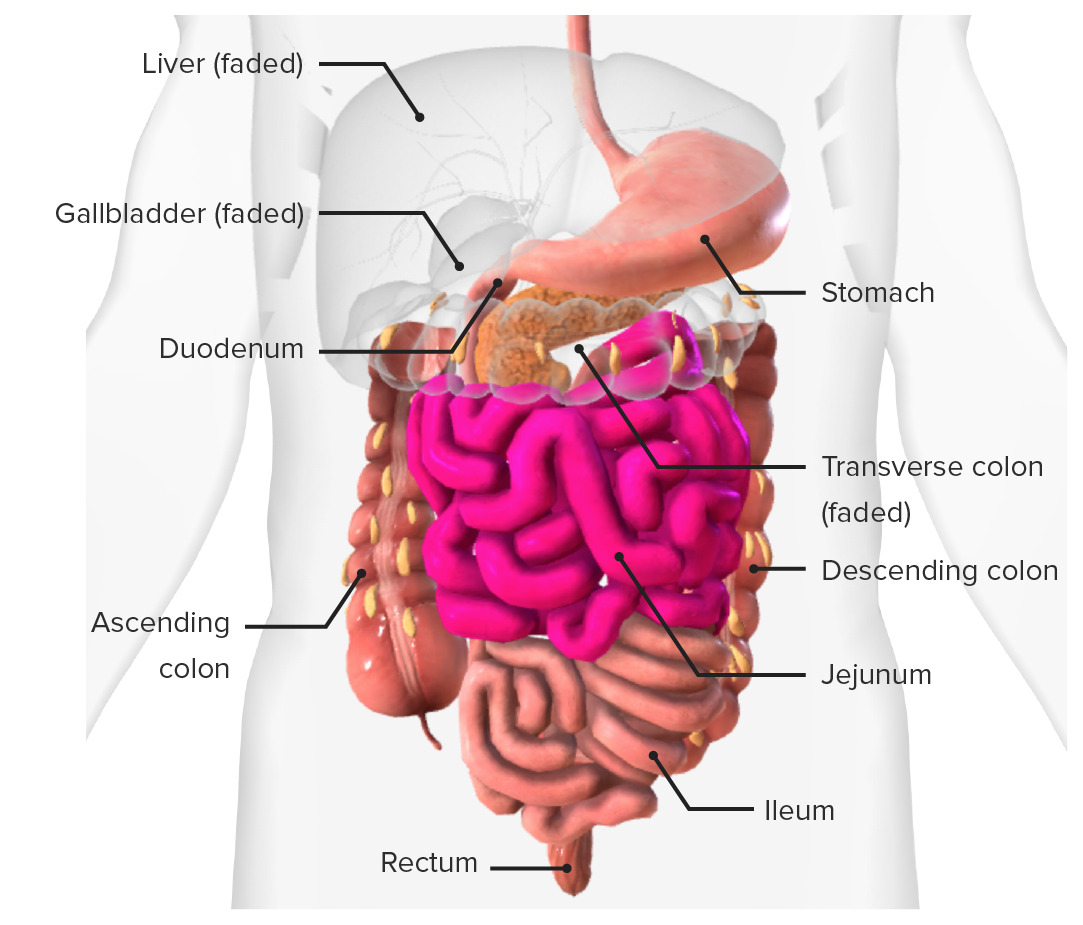
00:01 So let's have a look at the small intestine in situ within the abdomen if we were to remove the skin. So obviously we have the skin here over the anterolateral abdominal wall. The first thing we would encounter if we were to remove the skin and the muscle layers from the anterolateral abdominal wall and we entered into the abdomen would be a fatty apron. 00:23 This fatty apron is called the greater omentum and it's hanging down from the greater curvature of the stomach. Again, in some people it can actually be tucked up and actually adhere it to even the abdominal wall or various organs as a result of some localized infection. But classically and what you will see in many textbooks and many videos like this is a greater omentum actually drapes down from the greater curvature of the stomach and from the transverse colon of the large intestine and it covers all of the small intestine like an apron. If that were to reflect or that will be removed, you would see the small intestine is fined within the U shape or the inverted U shape of the large intestine. So here is the small intestine situated within the inverted U shape of the large intestine. You take away the transverse colon of the large intestine and you can start to see some organs which are deeper to it. Here we can see the stomach giving rise to the duodenum and then the duodenum, the C-shaped part of the tube running around the head of the pancreas up to the duodenojejunal junction. 01:32 Here we can see the stomach, we can see the pancreas sitting underneath the stomach, and the duodenum, the C-shape of the duodenum as described previously coursing around the head and the uncinate process of the pancreas then giving rise to the jejunum. The duodenojejunal junction is where this occurs. The jejunum then generally and it has this kind of torturous root, but is very much leaving from the upper left quadrant on its travels all the way down to the lower right quadrant. And about half way through that journey it transitions into the ileum. And we'll look at the characteristic differences of the jejunum and ileum later on. But the jejunum transitions into the ileum and then the ileum then goes and joins the cecum at the ileocecal junction. So here we can see the ileum, the terminal ileum, and the cecum. The cecum being this dilated sac-like structure at the beginning of the large intestine, and here at the ileocecal junction we have the ileum joining the cecum.
About the Lecture
The lecture In Situ View of the Small Intestine by James Pickering, PhD is from the course Anatomy of the Small Intestine.
Included Quiz Questions
Which portion of the intestine follows the duodenum directly?
- Jejunum
- Ileum
- Ascending colon
- Transverse colon
In which quadrant of the abdomen does the jejunum originate?
- Upper left
- Upper right
- Lower left
- Lower right
Which structure suspends the small intestine from the posterior abdominal wall?
- Mesentery
- Ligament
- Greater omentum
- Lesser omentum
- Fat
Which structure becomes continuous with the cecum distal to the ileocecal junction?
- Ascending colon
- Sigmoid colon
- Descending colon
- Rectum
Author of lecture In Situ View of the Small Intestine

James Pickering, PhD
Customer reviews
5,0 of 5 stars
| 5 Stars |
|
5 |
| 4 Stars |
|
0 |
| 3 Stars |
|
0 |
| 2 Stars |
|
0 |
| 1 Star |
|
0 |
