Playlist
Show Playlist
Hide Playlist
In Situ View of the Pancreas

-
Slides In Situ View of the Pancreas.pdf
-
Download Lecture Overview
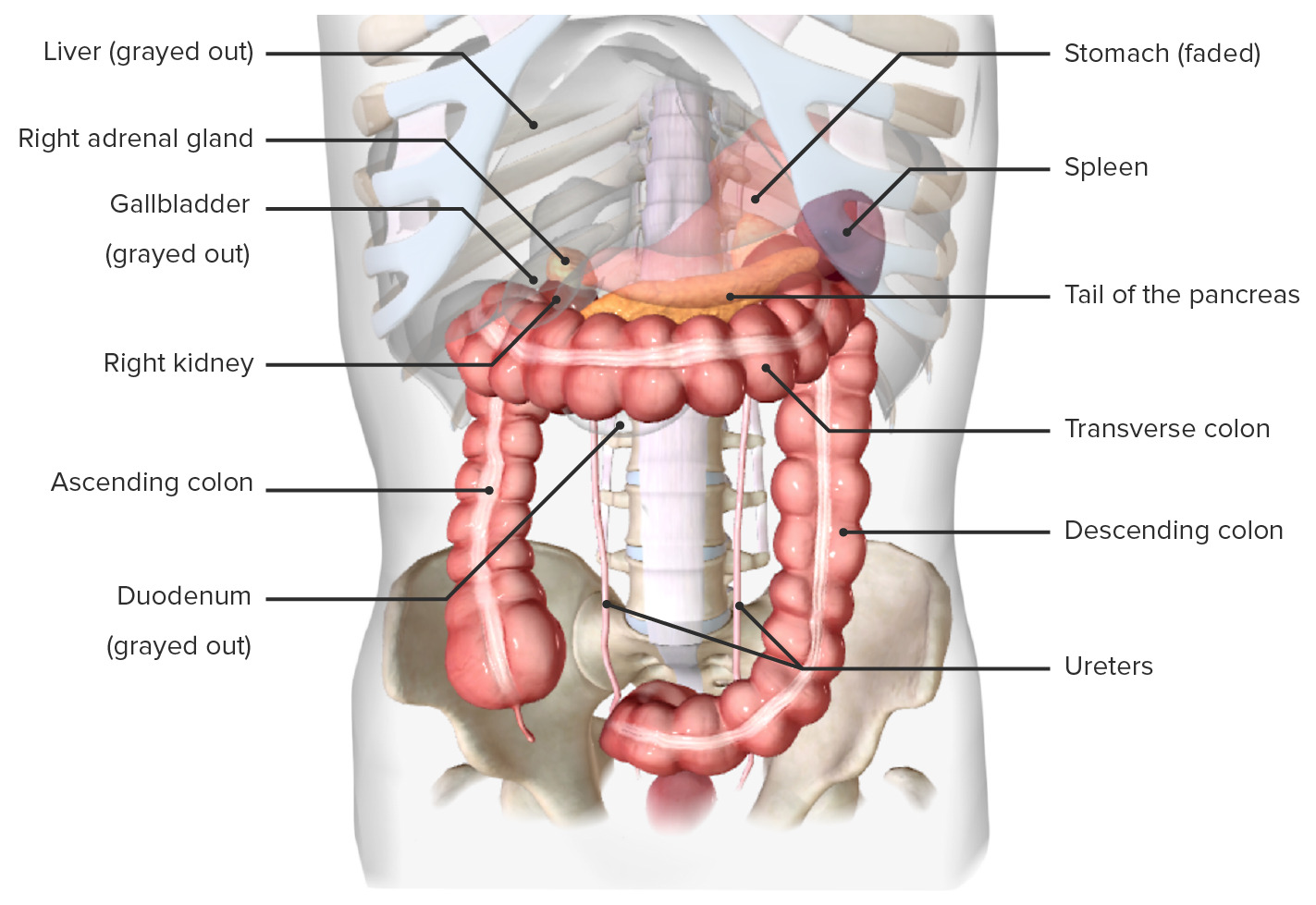
00:01 Now, let's have a look at the Pancreas In Situ with all of its neighboring organs surrounding it. 00:07 So here we have another typical picture of the abdomen. 00:10 We can see various structures such as the liver, the gallbladder, the transverse colon. 00:15 And we've got the jejunum, and the ileum. 00:17 All of these structures are sitting anterior to the pancreas. 00:21 If we were to remove those structures from the view, we can actually start to see the stomach is indicated and the duodenum. 00:28 So we can see the stomach giving rise to that C shaped duodenum. 00:32 And here tucked up on the left hand side in the upper left quadrant, we can see the spleen. 00:38 If we remove the stomach, we can start to see some other important structures. 00:43 So here we have the pancreas. 00:44 The pancreas is a retroperitoneal organ. 00:48 So it's sitting on the posterior abdominal wall with a layer of peritoneum sitting over it. 00:54 The exception is as the pancreas runs towards the hilum of the spleen, it assumes a covering of peritoneal and is actually intraperitoneal. 01:05 That's because the spleen has a peritoneal covering around it as well, which we'll come to in a moment. 01:11 So here we can see the peritoneal ligament, the splenorenal ligament, that's running towards the spleen, taking with it part of the tail of the pancreas. 01:24 We can also see the transverse mesocolon is coming off from the main kind of body and neck region of the pancreas. 01:31 So we have the transverse mesocolon, which goes towards the transverse colon. 01:35 And here we can see the head of the pancreas sitting within the concavity of the duodenum. 01:41 The neck of the pancreas is straddling over both the inferior vena cava and the aorta. 01:47 And we can see coming between the neck and the uncinate process of the pancreas. 01:51 We can see emerging so it sits anterior to the uncinate process and the duodenum, the superior mesenteric vessels. 02:00 So these are coming from posterior to the neck, they're emerging through the space between the neck, and the uncinate process, the C anterior over the duodenum. 02:11 Here we can see highlighted the body of the pancreas, and how that is associated with the aorta. 02:17 This region is around about the second, and the first lumbar vertebrae. 02:21 But again, this location can change from person to person, depending on the size of the stomach, depending on the location of the liver. 02:28 That can mean some of these organs do move around. 02:31 The tail of the pancreas, then heads towards the hilum of the spleen. 02:35 And we can see the tail of the pancreas, they're heading towards the spleen. 02:40 If we were to remove the pancreas, we can start to see sitting underneath it a lot clearer. 02:45 We have the right kidney, the hilum of the right kidney positioned underneath the right aspect of the head of the pancreas. 02:52 So we can see that on the right hand side. 02:55 We can also see coming down from the liver. 02:57 The common bile duct is that had run towards the second part of the duodenum. 03:01 And here, we can see the inferior vena cava much more clearly. 03:06 Running up on the underside of the neck of the pancreas, we see the hepatic portal vein. 03:11 We'll talk about the formation of the portal vein in a later video. 03:15 But that is receiving all of the blood from the gastrointestinal tract, and it passes underneath the pancreas to then join with the bile duct and the common hepatic artery to pass into the liver. 03:27 Here again, we can see the abdominal aorta sitting underneath the neck and the body of the pancreas. 03:34 Running alongside the posterior surface of the neck, body, and tail destined towards the spleen is the splenic artery and vein which we can see there. 03:44 And as it causes along that direction, the left kidney will be posterior to it.
About the Lecture
The lecture In Situ View of the Pancreas by James Pickering, PhD is from the course Anatomy of the Pancreas and Spleen.
Included Quiz Questions
Which structure runs posterior to the neck of the pancreas?
- Superior mesenteric artery and vein
- Inferior mesenteric artery
- Inferior mesenteric vein
- Splenorenal ligament
- Jejunum
In which structure does the tail of the pancreas run?
- Splenorenal ligament
- Phrenicocolic ligament
- Gastrosplenic ligament
- Transverse mesocolon
- Greater omentum
Which 2 veins unite to form the hepatic portal vein?
- Superior mesenteric and splenic
- Superior mesenteric and gastric
- Inferior mesenteric and gastric
- Inferior vena cava and splenic
- Gastroduodenal and splenic
Author of lecture In Situ View of the Pancreas

James Pickering, PhD
Customer reviews
5,0 of 5 stars
| 5 Stars |
|
5 |
| 4 Stars |
|
0 |
| 3 Stars |
|
0 |
| 2 Stars |
|
0 |
| 1 Star |
|
0 |
