Playlist
Show Playlist
Hide Playlist
In Situ View of the Large Intestine

-
Slides In Situ View of the Large Intestine.pdf
-
Download Lecture Overview
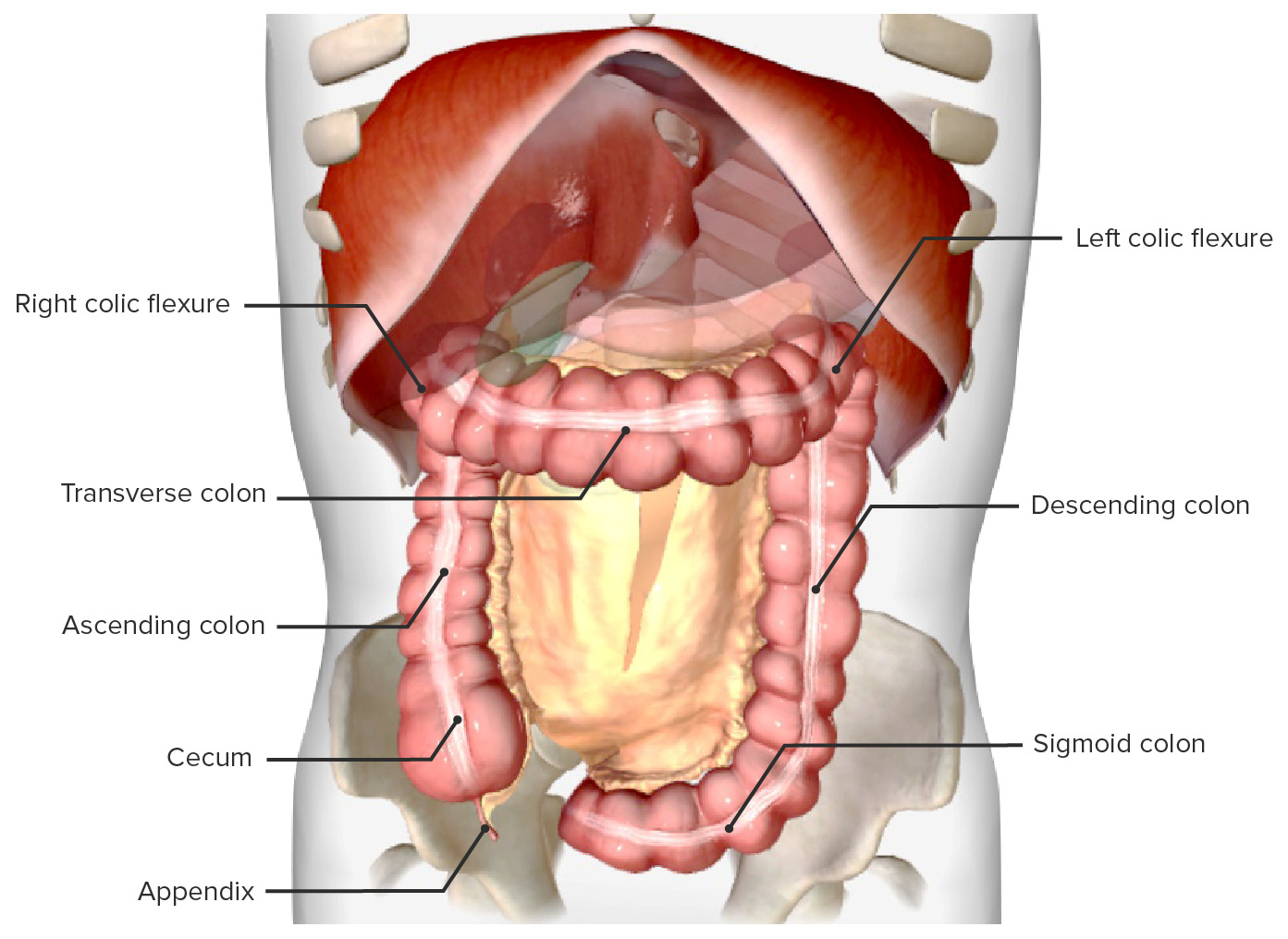
00:01 So if we then have a look at the large intestine in situ within the body, we can see here we have the outer layer of abdominal skin and then we remove the skin and we can see the anterolateral abdominal wall. Here's the rectus sheath, the aponeurosis here of external oblique. We remove that and then typically as seen in previous videos we see the greater omentum. And we'll come back to the greater omentum in the peritoneum video later on. We remove the greater omentum which is hanging down from parts of the transverse colon. And here we can start seeing the relationship of the cecum to various neighboring organs. So here we got the ileocecal junction, the continuation of the ileum into the large intestine, and it's really sitting on top of the right psoas muscle, the right iliacus muscle situated within the right lower quadrant of the abdomen. And here we can see the ileum that's feeding into it. The anterior abdominal wall obviously will be laying across this and actually the cecum aspect of the large intestine is really quite superficial so it's quite close to the surface of the skin and we'll come back to that when we look at McBurney's point in appendicitis in a moment or two. Here we can see the ascending colon. Again, it's running up and parallel with the right psoas major muscle and here is the iliolumbar ligament as well just for some relations with some neighboring structures. The right quadratus lumborum muscle is deep to the ascending colon and if we were to move the ascending colon and to the way you would see this muscle. Right transverse abdominus muscle crepes around the anterolateral aspects of the colon. And again, you can see the jejunum and ileum filling this space that is between the ascending transverse and descending colon. Again, the anterior abdominal wall would be directly above it. Here we got the hepatic flexure. The hepatic flexure is the turning point where the ascending colon on the right side of the abdomen then goes across to the left side and as the name indicates the hepatic flexure is associated with the liver. Here, most positively though we can see the right kidney and here we can see the undersurface or the inferior surface of the liver where the hepatic flexure is situated. The transverse colon then moves across to the left hand side of the liver. We can again see it touching the right kidney. We can see it running over the 2nd part of the duodenum. It also runs over the pancreas and here we've got the left kidney. Again, just sitting more anterior and below the transverse colon we find the jejunum. We can add the liver to the diagram as well, which is sitting over the top of the transverse colon and sometimes you really do need to lift the liver up and move the greater omentum and push the jejunum out of the way to really see this. But in there typically are for you to see the relationship with the transverse colon here. We got the liver, we got the gallbladder and deep to it we got the pancreas, etc. as we saw. An important structure that's connected to the transverse colon by way of the greater omentum is the greater curvature of the stomach and we'll come back to that when we talk about the greater omentum which we can see here, this fatty tissue, really a remnant of kind of peritoneal foldings and moving of the gastrointestinal tract, and we'll talk about in the peritoneum topic later on. Here we can see the splenic flexure of the colon, of the large intestine where the transverse colon then becomes the descending colon up by the spleen. So we can see the splenic flexure there. It's very much adjacent to the tail of the pancreas which runs towards the spleen. As you remember, we can see the left kidney is in this location as well and there is that colic impression on the spleen as the junction of the transverse colon and the descending colon run towards this upper left quadrant of the abdomen where the spleen is located. As we look at the descending colon, we've got very similar structures to what we had on the right hand side. Here on the left we've got the posterior abdominal wall and the various muscles that are loaded to. This will be the same as what's on the right hand side and here we have the small intestines, the jejunum and the ileum once again. The anterior abdominal wall again is going to be quite close to the surface of the descending colon and then we move into the sigmoid colon. Again, it's next to the various musculatures or iliopsoas here running down on the left pelvic wall. And we can also see we've got some very important blood vessels within this space as well. So the sigmoid colon is running over the iliac vessels. 04:22 Here, we can see part of internal and external iliac vessels and we've got various other tubes as in the vas deferens or the ductus deferens and also an important nerve, the obturator nerve, that's passing within this region as well. Running behind or posterior to the sigmoid colon here on the left hand side, we got the left ureter and again as I mentioned we got some important iliac blood vessels, both the external and the internal iliac vessels. Deep again to those is the piriformis muscle which is really lining the wall of the pelvis and that's an important muscle that passes out towards the hip. Most posteriorly again, the back of this space we've got the sacrum which is forming the posterior wall of the pelvis. Most anteriorly and sitting in front of the sigmoid colon we do have the bladder and obviously in the female specimen not shown in this picture there will be the uterus that sits between the rectum and the sigmoid colon becoming the rectum then moving anteriorly you'd have the uterus and then you'd have the bladder. Again, situated within this space we've got the jejunum and really the ileum that droops down into the pelvis and can sometimes obscure the sigmoid colon as it dives down to exit the pelvic floor as the rectum and then the anus. And there we have the small intestines. The rectum is an important continuation of the sigmoid colon and the rectum is that part of the gastrointestinal tract that leaves the pelvic floor to really go to the perineum where we have the anus. And here we can see the pelvic floor and the rectum passing to it. Again, directly posterior to the rectum we would have the sacrum and anterior to the rectum we would have the bladder. In this male specimen, we've got the bladder. In the female, you also have the uterus residing between the rectum here and the bladder. And again, in the male specimen here we can have the prostate to the base of the bladder.
About the Lecture
The lecture In Situ View of the Large Intestine by James Pickering, PhD is from the course Anatomy of the Large Intestine.
Included Quiz Questions
In which abdominal region does the large intestine begin?
- Right inguinal
- Left inguinal
- Right lumbar
- Left lumbar
Which 2 parts of the large intestine are suspended by mesentery?
- Transverse and sigmoid
- Transverse and ascending
- Transverse and descending
- Transverse and cecum
Author of lecture In Situ View of the Large Intestine

James Pickering, PhD
Customer reviews
5,0 of 5 stars
| 5 Stars |
|
5 |
| 4 Stars |
|
0 |
| 3 Stars |
|
0 |
| 2 Stars |
|
0 |
| 1 Star |
|
0 |
