Playlist
Show Playlist
Hide Playlist
Immunofluorescence Diagnosis in Vesiculobullous Dermatitis

-
Slides Dermatology Inflammatory Skin Diseases.pdf
-
Download Lecture Overview
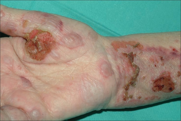
00:01 Here, we’ll take a look at a very important diagnostic image or images in which I will be comparing pemphigus vulgaris to bullous pemphigoid. 00:11 You want to pay a special attention to this page and to the images because this is the one in which it’s going to summarize everything that you need to know between the differences between the two diseases. 00:24 First, in general, it’s called vesiculobullous dermatitis. 00:28 But in other words, it’s a fact that you’re forming these blisters, either due to pemphigus vulgaris or bullous pemphigoid. 00:36 And the diagnostic tool that we’re using here is called immunofluorescence. 00:40 The last time we’ve seen the term or the diagnostic tool immunofluorescence was when we’re dealing with a type 3 hypersensitivity in renal pathology in which we were then identifying your immune complexes, or maybe perhaps your immunoglobulins in the basal membrane, right? For example, if you find immune complex in your basement membrane, your glomerular basement membrane, we call that lumpy bumpy our granular pattern of immunofluorescence. 01:10 So are we clear? Whereas, if it’s Goodpasture, then that IgG that was found in the basement membrane was of a linear pattern, all right? Now, with the premise in mind, what are we looking for? Immunoglobulins. 01:27 Now, the picture on the left, take a look at these immunoglobulins. 01:30 Beautifully highlighted at the dermoepidermal junction. 01:36 Stop and think and then answer. 01:39 We’re at the dermoepidermal junction. 01:41 The first question, what bridge is this? What’s the name of this protein or bridge that keeps the anchor between the dermoepidermal junction? Hemi-desmosome Good. 01:58 Hemi-desmosome is going to appear immunofluorescent, that makes no sense, because it’s not immunoglobulin. 02:05 Okay. 02:06 So, that means now, we have a pathology because the dermoepidermal junction is beautifully delineated. 02:13 You see that? And so therefore, an IgG is attacking the hemi-desmosome. 02:19 Diagnosis, please? Bullous pemphigoid, very good. 02:24 Tell me about the blister. 02:26 Tight or ruptured? Tight. 02:29 Large or small? Large. 02:32 Superficial or deeper? Deeper. 02:36 Bullous pemphigoid. 02:39 What about the right? On the right, you’ll notice that there’s absolutely no linear pattern of your immunofluorescence. 02:48 And specifically, it is the IgG which is then attacking the bridge between or the protein between your keratinocytes, right? And so therefore, this is referred to as being your desmosome or desmoglein proteins. 03:03 If this is being attacked by IgG, then what happens to your skin? It comes off, and we call this acantholysis, clear? The one the left, bullous pemphigoid. 03:17 On the right, pemphigus vulgaris. 03:20 Which one has an increased risk of mortality? The vulgar one, the pemphigus vulgaris. 03:25 Both are type 2 hypersensitivities, and we talked about which proteins are being attacked.
About the Lecture
The lecture Immunofluorescence Diagnosis in Vesiculobullous Dermatitis by Carlo Raj, MD is from the course Inflammatory Skin Diseases.
Included Quiz Questions
Which of the following patterns of immunofluorescence is most suggestive of bullous pemphigoid?
- Linear IgG staining at the dermal-epidermal junction
- Bright linear deposits of immunoglobulin G along the glomerular basement membranes
- Lumpy-bumpy granular deposits of IgG
- Intraepidermal deposition of IgG
- Intercellular deposition of IgG throughout the dermis
Damage to what structure is primarily seen in patients with pemphigus vulgaris?
- Desmosomes
- Hemidesmosomes
- Melanocytes
- Adipose tissue
- Keratohyalin granules
Author of lecture Immunofluorescence Diagnosis in Vesiculobullous Dermatitis

Carlo Raj, MD
Customer reviews
5,0 of 5 stars
| 5 Stars |
|
5 |
| 4 Stars |
|
0 |
| 3 Stars |
|
0 |
| 2 Stars |
|
0 |
| 1 Star |
|
0 |
