Playlist
Show Playlist
Hide Playlist
How to Approach Spinal Cord Pathology (Micro)
-
Slides Diseases of the Spinal Cord.pdf
-
Download Lecture Overview
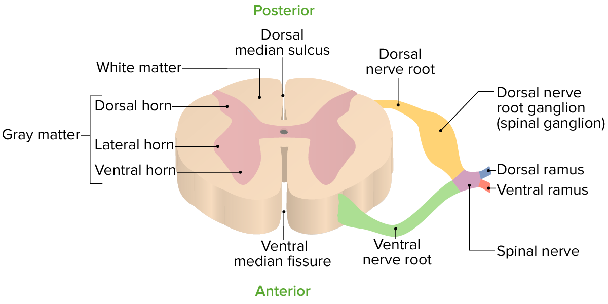
00:00 So let's look a little bit closer at how we approach various types of spinal cord pathology and we're going to walk through those 3 localizations in a patient who presents with findings concerning for myelopathy. We want to localize that to the extradural space, the intradural space, or the intramedullary space. Here we see a schematic of extradural pathology, and that is pathology outside the dura. This may be in the epidural venous plexus as you see this large red lesion here or arising from the vertebral body and compressing the spinal cord backwards. There's a couple of features that we see in patients presenting with this type of pathology. First of all, we see pain. 00:40 Back pain is really common from extradural pathology. The spinal cord itself doesn't actually feel pain. It just carries all the sensory nerves that carry perception and awareness of a painful stimulus. The pain fibers live in the dura and so extradural pathology that compresses the dura will present with pain and back pain is a red flag for potentially an extradural pathology. Frequently, we see extradural compression that is asymmetric or eccentric and you can see that here. The compression of the spinal cord is coming from laterally in to medially and so it may present with asymmetric onset of symptoms. And then finally, we see that this compressive pathology starts from the outside of the cord and compresses in. And in a few minutes, we'll look at the homunculus of the spinal cord, but the fibers that travel around the outside typically innervate and come from the legs and so we see leg symptoms first and the fibers that are on the inside of the spinal cord carry information about the arms and so we see arm or upper extremity pathology second. In that onset of leg symptoms followed by progressive difficulty with arm symptoms is suggestive of an extradural process. How about the intradural space? What does that look like? Well, here we see a schematic of a typical intradural extramedullary process, that means outside the spinal cord. Things like meningiomas and peripheral nerve sheath tumors and those sorts of pathology. This is often eccentric again compressing the spinal cord on one side more than the other and patients may present with asymmetric symptoms. 02:13 We can see pain but it typically develops later in the course of this presentation as the dura is slowly compressed and we often see lower motor neuron findings at the level of the lesion and we can see upper motor neuron findings below the level of this lesion. 02:32 And then lastly, we can see intramedullary pathology and that is pathology within the spinal cord itself and we're going to talk through some of the typical intramedullary syndromes that are important for you to know. In general, these are things that begin and develop within the spinal cord and expand the spinal cord or result in atrophy and contraction and reduction in tissue over time. Often, we don't see pain from intramedullary pathology because the dura is not compressed and pain is a feature of dural compression. We can see early bowel and bladder dysfunction and that should be suggestive of an intramedullary process. Let's look a little bit more at the somatotopy, the organization of the spinal cord and think about how this can help guide us in our examination of patients. We know the spinal cord carries motor information which descends in the white matter tracts and then exit out the individual segments of the spinal cord. The spinal cord also carries ascending sensory information up the white matter sensory tracts and then up into the brainstem and the thalamus to relay with the brain. When we think about those descending tracts, here you can see in this schematic an example of the lateral corticospinal tract. The corticospinal tract carries voluntary motor information, helps us to move our arms and legs when we want to and when we think about it. The lateral corticospinal tract carries about 85% of the motor information and the motor fibers going to the limbs, into the body. 04:02 This is the main voluntary motor control. It controls upper extremity motor symptoms and nervous innervation and lower extremity motor information. Pathways that are more lateral carry information for the legs and you can see the sacrum followed by the lumbar fibers and the thoracic fibers and the cervical fibers. Information and motor information, motor fibers for the arms are on the medial aspect within the spinal cord. And so again, externally compressive processes initially compress the nerves that are going to the lower extremities so we see lower extremity symptoms first followed by upper extremity symptoms. There there's also the ventral corticospinal tract which carries about 15% of the voluntary motor fibers. Let's look a little bit more at the sensory white matter tracts. We have 2 major sensory tracts in the spinal cord. The first is the dorsal column medial lemniscal system. This carries information for vibration, proprioception, and some deep touch. And again, you can see the somatotopic organization with the sacral fibers medial, lumbar, thoracic, and cervical fiber is a little bit more lateral. There is also the lateral spinal thalamic tract which carries information for pain and temperature and again you can see that somatotopic organization with lower extremity sensory information lateral and cervical sensory information carried medial. There is a ventral spinothalamic tract which carries information about light touch.
About the Lecture
The lecture How to Approach Spinal Cord Pathology (Micro) by Roy Strowd, MD is from the course Diseases of the Spinal Cord.
Included Quiz Questions
In patients with extradural myelopathy, symptoms begin in the lower extremities and progressively involve the upper extremities. Why does this occur?
- The fibers in the periphery of the spinal cord innervate and come from the legs, and the fibers deeper inside the spinal cord carry information about the arms.
- The fibers in the periphery of the spinal cord carry information about the arms, and the fibers deeper inside the spinal cord carry information about the legs.
- The fibers that innervate the legs are longer than those that innervate the arms.
- The fibers that innervate the legs are shorter than those that innervate the arms.
- The arms are closer to the brain.
Which of the following spinal cord tracts is the main descending tract that controls MOST of the voluntary motor information?
- Lateral corticospinal tract
- Dorsal column medial lemniscal tract
- Ventral corticospinal tract
- Spinothalamic tract
- Medial lemniscus
What information is carried by the dorsal columns?
- Proprioception and vibration sense
- Pain and temperature
- Pressure and pain
- Motor function
- Postural movements from visual stimuli
Which of the following represents the somatotopic organization of the dorsal columns (sensory information) from medial to lateral?
- Sacral, lumbar, thoracic, cervical
- Sacral, thoracic, lumbar, cervical
- Cervical, thoracic, lumbar, sacral
- Cervical, lumbar, thoracic, sacral
- Lumbar, thoracic, cervical, cranial
Customer reviews
5,0 of 5 stars
| 5 Stars |
|
5 |
| 4 Stars |
|
0 |
| 3 Stars |
|
0 |
| 2 Stars |
|
0 |
| 1 Star |
|
0 |


