Playlist
Show Playlist
Hide Playlist
Frontal Bone

-
Slides Anatomy Skull.pdf
-
Download Lecture Overview
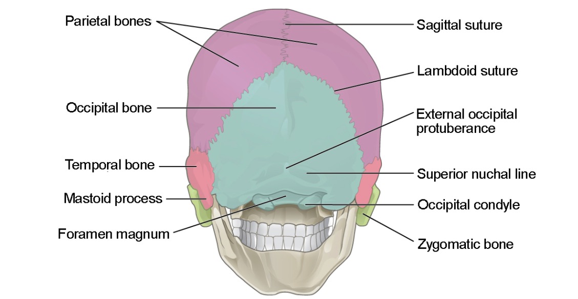
00:01 Let us now move on and discuss some of these bones in more detail. 00:06 The first bone that we will discuss is the frontal bone. 00:10 The frontal bone is the anteriormost bone of a neural cranium. 00:15 It is shell shaped and corresponds to the first frontal region of the face more commonly known as the forehead or the fronts. 00:26 Additionally, the frontal bone is said to have several different surfaces or parts, the orbital, the nasal, the squamous and although technically part of the squamous surface, we can also differentiate the zygomatic process. 00:45 The squamous part is the largest surface of the frontal bone. 00:49 This is specifically the portion that corresponds to the forehead. 00:53 There are several important bony landmarks located on the squamous surface of the frontal bone. 01:00 The first of which is the frontal tuber or tuberosity. 01:04 This landmark is located bilaterally approximately three centimeters above the eyebrow and it is a moderate prominence to which muscles and connective tissues can attach. 01:16 Below the frontal tubers and separated by a shallow groove are located two curved elevations known as the superciliary arches. 01:27 The superciliary arches are medially prominent inner joined together at the median elevated cranial metric point known as the glabella. 01:37 As a side note in certain individuals, a persistent line or suture extending from the glabella superiorly can be seen on radiographs. 01:47 This line represents the fusion side of the frontal bones embryologically and if present, it is referred to as the metopic suture. 01:56 Below this arch is located the supraorbital margin. 02:01 The margin acts as a boundary between the squamous and the orbital surfaces of the frontal lobe. 02:08 On the medial end of this margin is located a supraorbital foramen which permits the passage of the supraorbital nerve and vessels. 02:19 Medial to this foramen lies the frontal notch which can only be found in approximately 50% of skulls and permits the passage of the supratrochlear nerve and vessels. 02:33 And lastly for this sectio,n let's discuss the articulations of the squamous part of the frontal bone. 02:39 First, the lateral end of the supraorbital margin tapers sharply. 02:44 It ends in a prominent zygomatic process that articulates with the frontal process of the zygomatic bone. 02:52 Also from the zygomatic process, a curved posterosuperior line arises which divides into superior and inferior temporal lines. 03:02 What more, the area inferior to these lines is known as the temporal surface of the frontal bone, and it contributes to the formation of the anterior part of the temporal fossa. 03:13 Second, the posterior margin which articulates with the paired parietal bones, and then third, the inferior margin that articulates with the greater wing of the sphenoid. 03:25 And now we come to the last part of the frontal bone, the orbital part. 03:29 There are two landmarks in the orbital surface of the frontal bone that are of importance. 03:33 First of them is the fossa for the lacrimal gland. 03:36 This fossa is located on the anterolateral margin, the orbital plate and houses the lacrimal gland. 03:43 Second is the trochlear fovea. 03:45 This landmark is located on the anteromedial margin the orbital plate. 03:49 The trochlear fovea marks the attachment of the fibrocartilaginous trochlea through which the tendon of the superior oblique muscle passes. 03:57 Furthermore, another point to remember for future lectures is that the orbital part of the frontal bone makes a major contribution to the formation of the interior wall of the anterior cranial fossa. 04:09 But more on that in future lectures
About the Lecture
The lecture Frontal Bone by Craig Canby, PhD is from the course Head and Neck Anatomy with Dr. Canby.
Included Quiz Questions
The supraorbital margin is a boundary between which parts of the frontal bone?
- Squamous and orbital
- Nasal and orbital
- Zygomatic and orbital
- Nasal and squamous
- Squamous and zygomatic
What is the name of the frontal bone fusion line that can be visualized on imaging in some individuals?
- Metopic suture
- Atopic suture
- Extopic suture
- Frontal suture
- Midline suture
The trochlear fovea of the frontal bone attaches to the trochlea of which muscle?
- Superior oblique muscle
- Inferior oblique muscle
- Lateral rectus muscle
- Medial rectus muscle
- Inferior rectus muscle
Author of lecture Frontal Bone

Craig Canby, PhD
Customer reviews
5,0 of 5 stars
| 5 Stars |
|
5 |
| 4 Stars |
|
0 |
| 3 Stars |
|
0 |
| 2 Stars |
|
0 |
| 1 Star |
|
0 |
