Playlist
Show Playlist
Hide Playlist
Forearm in Cross-section – Anatomy of the Forearm

-
Slides 06 UpperLimbAnatomy Pickering.pdf
-
Download Lecture Overview
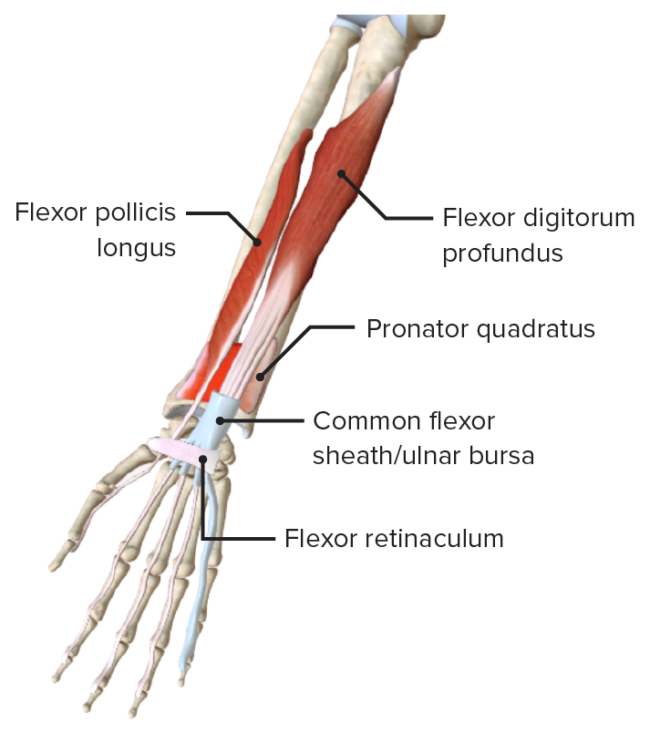
00:01 In this lecture, we’re going to look at the forearm. So we’ll start off by looking at the forearm in cross-section. We’ll look at the forearm fascia or the antebrachial fascia, and the various compartments that are formed. We’ll then look at these compartments, the anterior compartment and the posterior compartment. And in both of them, we’ll look at the various muscles, be a flexor, pronator or extensor muscles. We’ll look at various layers they formed, and also the various neurovascular relations. 00:34 So here we can see a cross-section through the forearm, showing the various muscles in their compartments, the interosseous membrane that’s running between the two bones, the radius and the ulna. And we can see that with this transverse section, we can divide the forearm into this anterior compartment here, and this posterior compartment. Remember, this is the inferior view and this is a right forearm. So we’re looking at it from below. We can see laterally, we have the radius, and medially, we have the ulna. These two bones are connected from the interosseous borders via the interosseous membrane. Radiating from these bones to the perimeter of the forearm, we have that intermuscular septae, and this is the continuation of the antebrachial fascia from the perimeter into the middle of the arm. So this intermuscular septae running across, and the interosseous membrane, forms the anterior compartment and the posterior compartment. Here, we can also see various blood vessels and nerves, so we can pick up the median nerve. We can see the ulna nerve and the ulnar artery. We can see the radial artery and the radial nerve as well. And we’ll look at this in more detail as we go through this lecture. The anterior compartment contains muscles that are ultimately going to flex the wrist. They’re also associated with pronation of the forearm. The posterior compartment contains muscles that are principally going to extend the wrist and also supinate the forearm. But there’s also some other movements that can occur, and whilst the majority of the muscles in the forearm act on the elbow, they also act on the radio-ulnar, the wrist, metacarpophalangeal and interphalangeal joints. And at these joints, there can be a whole series of movements created by the muscles within the forearm. So these muscles in the forearm can send long tendons that go and attach quite distally to the very distal phalanges of the digits. You can have flexion and extension. These can occur at the elbow joints, at the wrist joints, at the metacarpophalangeal, and interphalangeal joints. So, most of these joints will enable flexion and extension. At the radio-ulnar joints, we have pronation and supination, and we’ll look at some important muscles that do that. And also at the wrist joint and at the metacarpophalangeal joints, we can have adduction and abduction.
About the Lecture
The lecture Forearm in Cross-section – Anatomy of the Forearm by James Pickering, PhD is from the course Upper Limb Anatomy [Archive].
Included Quiz Questions
Which movements are performed by muscles of the anterior compartment of the forearm?
- Flexion and pronation
- Extension and supination
- Extension and pronation
- Flexion and supination
- Adduction
Author of lecture Forearm in Cross-section – Anatomy of the Forearm

James Pickering, PhD
Customer reviews
5,0 of 5 stars
| 5 Stars |
|
5 |
| 4 Stars |
|
0 |
| 3 Stars |
|
0 |
| 2 Stars |
|
0 |
| 1 Star |
|
0 |
