Playlist
Show Playlist
Hide Playlist
Filtration Barrier
-
Slides RenalClinicalAnatomy RenalPathology.pdf
-
Download Lecture Overview
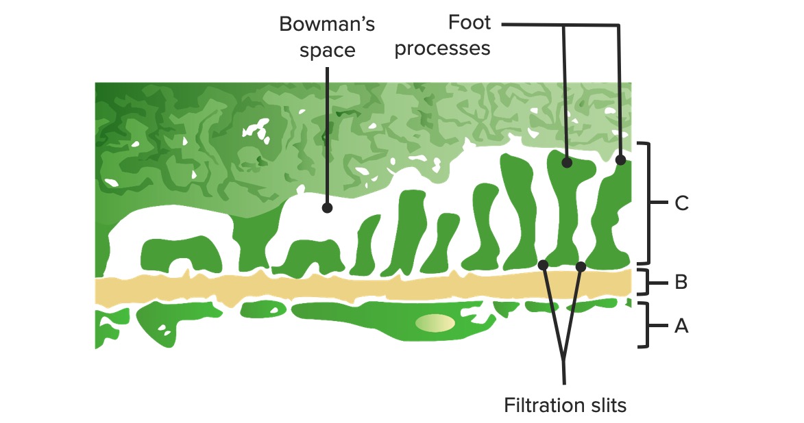
00:00 We begin with this slide as being quite important. Filtration barrier is what we'll set up and before you do any of that, I am going to give you steps that you want to take every single time. 00:12 When you see a picture like this, that is then representing electron microscopy. Now from a bird's eye of view, what exactly is occurring and why would you want to use such tools at your disposal? Well you know that the patient is demonstrating glomerular nephritis, how? Well as we go through the particular diseases obviously I will give signs and symptoms. Next, well what particular type of biopsy and imaging studies subsequently would you want to perform so that you can actually find the pathology? So upon biopsy, you might want to begin with light microscopy, which is not what is being shown here. But light microscopy is something that will give you a bird's eyeof view of the glomerulus and then say that you don't find your pathology then perhaps you go into a little bit detail and that, of course, brings us to our electron microscopy and that is where we are at this juncture. What I am going to show you first would be the normal electron microscopy of the exchange system or the filtration system of your fluid from the plasma. Close your eyes. You are in the tuft of capillaries, that's your plasma. The first barrier that you are going to hit is what? Is that an endothelial cell or an epithelial cell? It is an endothelial cell. Are we clear? Because it is lining of the blood vessel. I will show you what they are as you move through here, just I want to conceptualize things first. Underneath that endothelial cell is what? That is what we're saying here. So this would be the first paved road and by paved road, this is the first thing that you want to identify in electron microscopy so that you immediately get your bearings. The bearings here would be this paved road and this would then represent your filtration or your basement membrane. A couple of things about this basement membrane that you have learned in anatomy and then reinforce in physio and then we'll add some pathologies here include the fact that well and as far as chargers are concerned, it has a negative charge. It has to. Now, let me tell you the objective first so that you understand this. Albumin would be on the blood side, wouldn’t it? May I ask you a question? Do you want albumin to pass through? Simple question, but yet effective. No, you don't. If you don't want albumin to pass through, you should know the protein as a negative charge. We've talked about such things with the double helix of the DNA. Our protein has a negative charge in general. 02:43 In physics, when you have two likes and charges, what do they do? Not relationships. These are charges, when you have two likes, they repel. So, therefore, albumin has a negative charge, the basement membrane has a negative charge. Thus, what about that albumin? It is repelled. That is one form of defense. A second form would be well you know filtration is taking place. The pores, the fenestrations that exist that allow for filtration to take place. They are quite tiny. They have to be. So these pores and fenestrations that are quite tiny do not allow the large albumin proteins to pass through. Two major natural defense mechanisms or barriers that preclude albumin from passing through. If at any point in time, pathologically, there is abnormality with that pore getting larger or the negative charge has been lost, you have now introduced albumin into the Bowman space and you have a true pathology and we will walk through that. So now what we are seeing here, the blood side is what we will talk about in the fenestrations. In the fenestrations here then allow for filtration to take place. Now where the fenestration here is in reference? Now we are going to go step by step. On the right side of this barrier would be the endothelial cell. There would be the blood side. Stop there, identify. That is your blood side. In between the endothelial cells are technically called fenestrations and that you must understand. 04:19 Next, while you have that glomerular basement membrane and that glomerular basement membrane is then going to be made up of a negative charge. When we talked about the fenestrations being extremely, well small enough, large enough just right where electrolytes and small amino acids will pass through filtration. But please understand that large protein will not. 04:44 So it is a negatively charged. It has to be. Next on the side of the urine is where we are here. On the side of the urine well now we have left the blood vessel. Is that clear? When you’ve left the blood vessel and now you are dealing with a cell of a tissue, that cell of a tissue is then called an epithelial cell, isn't it? The epithelial cell and specifically take a look, this is the visceral epithelial cell. What does that mean? The inner layer, the visceral epithelial cell is then called a podocyte. I would know about the names. 05:25 Why do we call this a podocyte? What is the profession of podiatry? Who is a podiatrist? A "foot" doctor right. Oh! Look at your feet, I love them so much. Anyhow, that's the foot doctor, podiatrist. The point is this is a podocyte and so therefore what you will find are this foot processes. In between the foot processes are slit diaphragms and so therefore as your filtration now you tell me which way filtration on this electron microscopy? What are you doing when you filter? You are moving from the plasma into the Bowman space. On this picture is it left to right or is it right to left? Good. It is right to left. That is filtration. You are leaving the plasma on the right side, passing through the basement membrane, and then moving through your slit diaphragms of your podocyte. Everything that you are seeing here is perfectly normal. I need you to get a firm grasp of what we are looking at with electron microscopy. What is the first thing that you want to do? Paved road. From the paved road, you look for the feet, the foot processes that automatically puts you on the urine side. We are going to keep playing around with this and until you get this firmly implanted. Let's move on.
About the Lecture
The lecture Filtration Barrier by Carlo Raj, MD is from the course Renal Clinical Anatomy.
Included Quiz Questions
Which of the following depicts the correct sequence of the layers of the filtration barrier?
- Fenestrated endothelial cells, negatively charged glomerular basement membrane, and visceral epithelial cells
- Fenestrated epithelial cells, negatively charged glomerular basement membrane, and visceral endothelial cells
- Fenestrated endothelial cells, positively charged glomerular basement membrane, and visceral epithelial cells
- Fenestrated endothelial cells, negatively charged glomerular basement membrane, and parietal epithelial cells
- Fenestrated epithelial cells, negatively charged glomerular basement membrane, and visceral epithelial cells
What are the defense mechanisms that prevent the filtration of albumin from passing through the glomerulus?
- Tiny fenestrations of the endothelial cells and a negatively charged glomerular basement membrane
- Large fenestrations of the endothelial cells and a negatively charged glomerular basement membrane
- Tiny fenestrations of the endothelial cells and a positively charged glomerular basement membrane
- Tiny fenestrations of the epithelial cells and a negatively charged glomerular basement membrane
- Tiny fenestrations of the endothelial cells and negatively charged epithelial cells
Which of the following statements represents the correct match?
- Fenestrations in the endothelial cells and filtration slits in the epithelial cells
- Fenestrations in the epithelial cells and filtration slits in the endothelial cells
- Fenestrations in the podocytes and filtration slits in the endothelial cells
- Fenestrations in the endothelial cells and slit diaphragms in the glomerular basement membrane
- Fenestrations in the epithelial cells and slit diaphragms in the endothelial cells
Which of the following is not a component of the luminal side of the filtration barrier?
- Parietal epithelial cells
- Podocytes
- Slit diaphragms
- Visceral epithelial cells
- Foot processes
Customer reviews
4,0 of 5 stars
| 5 Stars |
|
0 |
| 4 Stars |
|
1 |
| 3 Stars |
|
0 |
| 2 Stars |
|
0 |
| 1 Star |
|
0 |
I believe the direction (left to right and right to left) needs to be reversed


