Playlist
Show Playlist
Hide Playlist
Femur
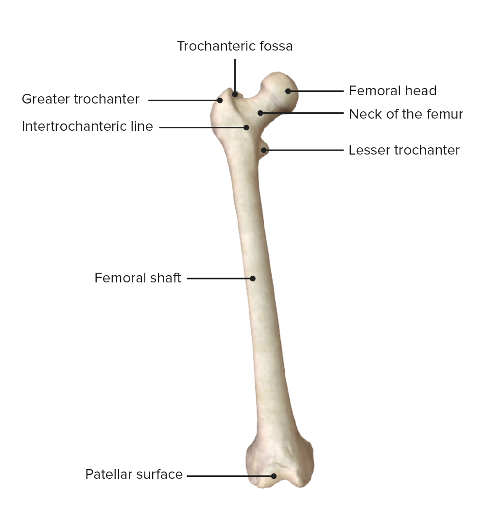
00:01 So now let's continue with the lower limb and start looking at the femur. 00:08 So the femur is a very long solid firm bone that is attaching the hip joint approximately to the knee joint distally and we can see we've got three broad areas to our femur here. 00:20 We've got the proximal end that forms the hip joints with the acetabulum of the pelvis. 00:25 We then have the long shaft and then we have the distal end of the femur which forms the knee joint with the tibia. 00:32 If we have a closer look at the proximal end, we can see we've got this very large dome-shaped head and we have various features associated with this proximal end. 00:42 So we have the head of the femur, which articulates with the acetabulum and that is connecting to the shaft of the femur by way of a neck. 00:50 And then as the neck becomes the shaft, we have these dilations, the swellings which really are the greater and the lesser trochanter has. 00:58 These are connected by what's known as intertrochanteric line. 01:02 And then there's a little lip that forms underneath the intertrochanteric line and that is the trochanteric fossa. 01:10 So we have a very dome-shaped head, that head is connected to the shaft of the femur by way of a neck. 01:17 Then as the shaft begins, we have the greater and the lesser trochanters. 01:21 These are bony protuberances that dilate and swell the beginning of the shaft of the femur to offer muscle attachment sites connecting those is an intertrochanteric line. 01:32 And then there is a trochanteric fossa that just sits underneath that lip of the line I just described. 01:39 So if we then have a closer look at the head, we can see that the head is articulating with the acetabulum of the hip bone, remembering the acetabulum is formed by those three bones of the hip, the pubis, the ischium, and the ilium. 01:53 And we can see together that forms the hip joints, you'll notice the depth of the acetabulum. 01:58 And the dome-shaped of the head is quite different to that of the shoulder joint which had a much shallower glenoid cavity and a much shallower head of the humerus. 02:09 That's obviously because the head of the femur articulating the acetabulum needs to be a lot more stable than that of the shoulder joints. 02:18 But that compromises movement, whereas for the shoulder, you kind of have a compromised by the amount of stability, you have to help give you much more movement. 02:29 If we then have a look at the head of the femur, we have this very kind of central fovea, this little opening at the very top of the head, and that contains a ligament, the ligament of the head of the femur or the round ligament of the femur. 02:41 And that helps to hold the head in position within the acetabulum. 02:46 If we don't have a look at the posterior surface of the same view, we see similar structures, we see the greater trochanter superiorly. 02:53 And that gives rise to the lesser trochanter by way of the intertrochanteric crest which we can see now on the posterior surface. 03:01 We had a line on the anterior surface now we've got a crest on the posterior surface. 03:06 And then if we move down into the shaft of the femur, we can have a look at both the anterior and the posterior surfaces. 03:14 On the anterior surface, we obviously have a lateral border, that's the opposite side of where the head of the femur is, the head of the femur is on the same side, obviously, as the medial border of the femur. 03:26 And then posteriorly we have really a kind of prominent sharp edge that arises posteriorly and that forms the posterior border of the femur. 03:35 This creates a number of surfaces. 03:37 So between those borders, we obviously have surfaces. 03:41 So between the lateral and the medial borders, we have the anterior surface. 03:46 And then where posteriorly, it elevated into this kind of sharp ridge which formed the posterior border that then creates the postural and medial and posterior lateral surface. 03:56 And these surfaces are important to bear in mind when we think about muscle attachments, which we'll come to later on. 04:03 So now let's have a look at the posterior surface in a little bit more detail. 04:07 Specifically that elevated reach that we saw on the posterior surface or really the posterior border we call that the linea aspera. 04:15 And here we can see is forming its medial lip, and here its lateral lip which gives those postural lateral surfaces, posteromedial surfaces on our femur. 04:26 Here we can see the pectineal line. 04:28 We can see a gluteal tuberosity. 04:30 We can also see the medial supracondylar line here and the lateral supracondylar line here as well. 04:38 Some important landmarks, which we don't need to worry about at the moment but they offer attachment sites for muscles. 04:43 So it's important that you can appreciate these bony landmarks on the various bones we discussed. 04:49 So immediate lip lateral lip converging together to form the linea aspera which then separates again as a supracondylar lines medial and lateral. 04:58 They go on to form the condyles of the femur. 05:01 And then more superiorly, we have the pectineal line and the gluteal tuberosity. 05:06 If we finish off looking most inferiorly as we head towards the knee, I mentioned we have the medial and lateral condyles that are forming from those supracondylar lines. 05:16 In between those two lines, we have the beginning of the poplar to surface and that really is beginning to demarcate the popliteal fossa which we'll come to later on. 05:27 So let's have a look at the distal end of the femur and this is looking at the anterior surface. 05:32 So you can very much see the patellar surface here. 05:35 The patellar surface is obviously where the patella is going to sit. 05:38 And that's important as it transmits the quadriceps tendon from the anterior thigh down onto the tibia. 05:45 So we've got the patellar surface. 05:47 And here we can see it's articulating with the flat surface superior surface of the tibia. 05:52 And here we're looking at the proximal end of the tibia. 05:55 So here is the distal end of the femur is articulating with the proximal end of the tibia. 06:00 And the patella is sitting between those two on its most anterior of aspects. 06:06 And these together form the knee joint. 06:08 Notice what's not in the knee joint here is the fibula, the fibula doesn't form part of the knee joint. 06:15 Let's have a look at the posterior surface now. 06:18 And we've got a couple of structures I mentioned a few moments ago, we've got the medial and lateral condyles. 06:24 Here we have the medial and lateral epicondyles, and they're really much coming from those supracondylar lines I mentioned. 06:32 On the most medial aspect, we have the adductor tubercle. 06:35 And in between the two condyles, we have the intercondylar fossa. 06:40 It's really important that you're just familiar with the names and the location of the structures at the moment. 06:45 It's not important to worry too much about them. 06:48 Because really, we need to remember them when we talk about muscle attachments. 06:52 And then we'll be talking about various muscles attaching from these bony points. 06:57 To finish off, we just have that red line that's appeared and that is really separating this intercondylar fossa from the popliteal soft surface we can see there in blue and that's the intercondylar line. 07:09 Now let's look at the articular surface of the distal end of the femur. 07:14 Here we got the patellar surface, that's where the patella runs up against. 07:18 And then we have the medial epicondyle and the medial condyles. 07:20 Now we're looking at it as if we're looking at the most distal end of the femur, we're looking at the articular surfaces especially where the medial and lateral condyle was at. 07:30 The lateral and the epicondyles are what are giving rise to those condyles as they emerge down from the shaft of the femur. 07:37 Now we can see in a different perspective, the actual space that's created by those two dilations of the condyles, the intercondylar fossa. 07:45 These are important because running within this space, we're going to have a series of ligaments, specifically the cruciate ligaments which helped to stabilize the knee joint as we are walking and moving.
About the Lecture
The lecture Femur by James Pickering, PhD is from the course Osteology and Surface Anatomy of the Lower Limbs.
Included Quiz Questions
Which part of the proximal femur is most inferior?
- Lesser trochanter
- Neck
- Head
- Greater trochanter
- Trochanteric fossa
What is contained within the central fovea of the head of the femur?
- Round ligament of the femur
- Acetabular ligament
- Trochanteric ligament
- Trapezius
- Oblique ligament
What is the longitudinal crest on the posterior surface of the femoral shaft?
- Linea aspera
- Pectinael line
- Gluteal tuberosity
- Medial supracondylar line
- Lateral supracondylar line
What separates the intercondylar fossa and the popliteal surface?
- Intercondylar line
- Lateral epicondyle
- Lateral condyle
- Medial condyle
- Adductor tubercle
Customer reviews
5,0 of 5 stars
| 5 Stars |
|
5 |
| 4 Stars |
|
0 |
| 3 Stars |
|
0 |
| 2 Stars |
|
0 |
| 1 Star |
|
0 |


