Playlist
Show Playlist
Hide Playlist
Female Perineum and Vulva

-
Slides Female Perineum and Vulva.pdf
-
Download Lecture Overview
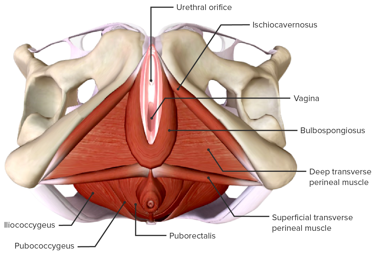
00:01 Now, let's turn our attention to the female perineum. 00:05 So here we're looking at the anterior aspect of the pelvis, which has just been tilted slightly posteriorly. 00:12 so we can see both the urogenital triangle and the anal triangle. 00:16 At the top of the screen again we can see the pubic symphysis. 00:19 Here we can see the perineal membrane has been added in to separate the superficial from the deep perineal pouch of the urogenital triangle. 00:27 And here we can see within the deep perineal pouch by removing the perineal membrane. 00:32 We can see some important blood vessels that have run from the pudendal canal and now entering into the urogenital triangle. 00:42 If we then add on to this region, the deep transverse perineal muscle then we can add in other important muscular structures like the urethra and external urethral sphincter. 00:51 And here we have the opening for the vagina as well. 00:54 The blood vessels like I said are going to be coming from the pudendal canal. 00:58 So we have the internal pudendal artery vein. 01:01 Will also have the pudendal nerve in this space as well. 01:04 And these have run out to the greater sciatic foramen, from the pelvis into the perineum. 01:09 Run along the shear pubic canal giving off branches to the anus. 01:13 But then they've carried on running forwards into the urogenital triangle of the perineum. 01:19 And here we can see them within the deep perineal pouch. 01:23 So now if we add in this perineal membrane when they're looking at the superficial perineal pouch here. 01:29 So this gray sheath is the perineal membrane. 01:32 And similar to the male these are analogous structures. 01:35 We have our external genitalia. 01:37 So laterally we have the crus of the clitoris. 01:40 Instead of having the crus that went on to form the penis, we now have the crus of the clitoris. 01:45 With the clitoris and the glands penis being analogous structures. 01:49 Here we have the body of the clitoris most distally from the crus sitting near the pubic symphysis. 01:55 And here we can see the bulb of the vestibule running up towards it. 02:00 The bulb of the vestibule is similar to the bulb with the penis. 02:04 But importantly, in the female, they have an additional opening which lies posterior to the urethra. 02:10 And the opening of the vagina means the bulb of the penis in the male has been split via that vaginal opening. 02:17 So we have the bulb of the vestibule that flanks either side of the vaginal opening or the vestibule. 02:25 So then here we can add on the muscle layer that we saw also in the male covering the bulb of the penis. 02:31 This time it's covering the bulb of the vestibule and this is the bulbospongiosus muscle also covering the crus of the clitoris we have the ischiocavernosus muscle. 02:40 And exactly the same process happens in the female as what happens in the male. 02:45 So, upon sexual arousal, the penis becomes erect as blood fills these erectile tissues. 02:51 Exactly the same process happens in the female but obviously there isn't as much of a substantial structure because the glans clitoris is that much smaller. 02:59 But the same process is happening. 03:02 During sexual arousal these erectile tissues will becoming good with blood. 03:06 And the muscles that surround these erectile tissues bulbospongiosus and the ischiocavernosus muscle will let restrict the venous return of blood from those erectile tissues maintaining the erection of the clitoris. 03:19 So as sexual arousal occurs, the clitoris will becoming gorged with blood and you'll have that clitoral erection. 03:27 The final muscle we can see here is the superficial transverse perineal muscle, which as it contracts helps to add support to the perineal membrane where these important structures are emanating from. 03:39 So let's have a look at the superficial features of the perineum in the female. 03:44 So here we can see some familiar landmarks. 03:46 We have the anus most posteriorly. 03:49 Within the anal triangle of the perineum. 03:51 And here we can see the perineal raphe that's running towards the vulva or the opening to the vagina. 03:58 Here we can see the labia majus which is uniting anteriorly and posteriorly as the commissure of the labia majora. 04:04 And here we can see lying medial to the labia majus so we have the labia minor. 04:09 We can see those two layers of skin that often fatfield helping to protect the opening of the rest of vestibule there. 04:16 Here we have the frenulum of the labia minora. 04:18 Here we have the prepuse of the clitoris. 04:20 Here we have the glans clitoris. 04:22 The prepuse is just to skin covering that lines over the glans clitoris. 04:27 Here we have the frenulum of the clitoris that's connecting it to the labia minus and here we have the external urethral orifice. 04:33 For the urethra to pass out of the urogenital triangle. 04:37 Most posterior to the urethra, we then have the vestibule of the vagina. 04:41 So it's important to remember that from anterior to posterior, the key landmarks are the clitoris, the urethra, the vagina, and then the anus. 04:50 And we can see those depicted on the surface of the female.
About the Lecture
The lecture Female Perineum and Vulva by James Pickering, PhD is from the course Perineum.
Included Quiz Questions
What is NOT a component of the external female genitalia?
- Perineal membrane
- Glans clitoris
- Crus of clitoris
- Bulb of vestibule
- Body of clitoris
What are the muscles within the female perineum? Select all that apply.
- Bulbospongiosus muscle
- Ischiocavernosus muscle
- Corpus cavernosum
- Deep transverse perineum
- Levator ani
What is NOT a superficial feature of the female perineum?
- Ischioanal fossa
- Labium majus
- Labium minus
- Perineal raphe
- Anterior commissure of the labia majora
Author of lecture Female Perineum and Vulva

James Pickering, PhD
Customer reviews
5,0 of 5 stars
| 5 Stars |
|
5 |
| 4 Stars |
|
0 |
| 3 Stars |
|
0 |
| 2 Stars |
|
0 |
| 1 Star |
|
0 |
