Playlist
Show Playlist
Hide Playlist
Extrinsic Tendons and Lumbricals
-
Slide Extrinsic Tendons Lumbricals.pdf
-
Download Lecture Overview
00:01 So, now, let's have a look at the actual muscles that form within the substance of the hand. 00:06 So, these kind of deep muscles associated with the metacarpals and the digits. 00:12 So, let's remind ourselves of some of the tendons that are intimately associated with them as well. 00:16 So, here, we have the flexor digitorum superficialis tendons and sitting deep to them, those profundus tendons as well, passing from the forearm into the hand, passing through the carpal tunnel, and then, entering this central compartment. 00:31 What we've done here is we've removed the palmar aponeurosis. 00:34 We've moved the muscles that supply - that are found within this region and we're just looking at that central compartment deep to the palmaris aponeurosis. 00:44 We can see the tendons and we can see some interosseus muscles there. 00:48 But these tendons are passing to the second through to the fifth phalanges of the second to fifth digits. 00:55 So, we can see them passing in a very specific orientation. 00:59 So, here, we can see most superficially the tendon of flexor digitorum superficialis. 01:06 And this is occurring at each of those four digits. 01:09 And what you can see sitting most superficially is that tendon of flexor digitorum superficialis. 01:16 But its attachment site is the middle phalanx. 01:19 So, you can actually see highlighted in blue how that tendon splits into two parts, attaching to the middle phalanx of the digit. 01:28 Here, we can see it's indicated as the lateral and medial surfaces of the middle phalanx. 01:33 But what you can also see is coming through that split is the tendon of flexor digitorum profundus. 01:40 So, if you look at the image on the left-hand side of the screen, flexor digitorum profundus tendon is sitting deep to superficialis. 01:48 Yet, its attachment site is the distal phalanx. So, the only way it can get to the distal phalanx is if it penetrates through the substance of flexor digitorum superficialis and it does this by passing through that split in flexor digitorum superficialis and then, going onto attach to the distal phalanx. So, this is how you have flexor digitorum superficialis attaching to the middle phalanx and flexor digitorum profundus tendon attaching to the distal phalanx. 02:21 The tendon passing through the split, the profundus tendon passing through the split of the superficialis tendon to go to the more distal phalanx. 02:32 It's a very important relationship that you should be familiar with. 02:37 Now, let's have a look at some other muscles in this space. And these are called lumbricals. 02:42 And these are intimately associated with those tendons we've spoken about. 02:46 We have four of these. The lateral two, Lumbricals I and II are unipennate, so, they have one muscle belly. Whereas Lumbricals III and IV are bipennate, they have two muscle bellies and you can see them by their origin here. 03:03 So, if we were to have a look at the unipennate muscles, the Lumbricals I and II, you can see these are coming from tendons flexor digitorum profundus. 03:14 So, these muscles actually originate from the tendon that we've spoken about beforehand and they pass all the way towards the distal phalanges, the phalanges of the hand. 03:26 So, here, we can see them passing from tendons of digitorum profundus muscle. 03:32 We can also see that the bipennate muscles, so, Lumbricals III and IV, these also come from the ulnar side of these tendons. 03:41 So, whereas you could see on the previous slide, all of these lumbrical muscles coming from the radial aspect, the ones with a bipennate, so, two heads, Lumbricals III and IV are also coming from the ulnar side of these tendons, only happens on III and IV which is why they are bipennate. 04:01 But these muscles pass towards the distal phalanges of the digits really by merging with the extensor expansions that we spoke about when we looked at the dorsal surface of the hand. 04:13 They don't go and attach to a specific phalanx, they attach to those extensor expansions of the digits around the hand. Here, we can see the innervation of these muscles. 04:24 So, here, we can see Lumbricals I and II, supplied by branches of the median nerve. 04:29 And here, we could see Lumbricals III and IV supplied by branches of the ulnar nerve. 04:35 So, I and II on the more lateral aspect supplied by the median nerve and Lumbricals III and IV on the more medial aspect supplied by the ulnar nerve. 04:45 So, if we look at the function of the lumbricals, then, their orientation, their position is really quite important which gives them a strange function in terms of movement of the fingers. 04:55 Here, we could see the lumbrical muscles passing across the metacarpo-phalangeal joint. 05:00 It passes anterior to that metacarpo-phalangeal joint. Meaning that when it contracts, it flexes that joint. 05:07 But as it moves to the side and attaches to the extensor expansion hoods, it actually attaches posterior to this interphalangeal joints. 05:18 And as it's attaching posterior to the interphalangeal joints, it actually works by extending those interphalangeal joints. 05:27 So, what we have by the lumbricals is really an extension and a flexion movement with bones that are very closely organized together. 05:34 So, the lumbrical muscle helps to flex the metacarpo phalangeal joint but extend the interphalangeal joints. 05:43 And that's an important difference you should try and remember.
About the Lecture
The lecture Extrinsic Tendons and Lumbricals by James Pickering, PhD is from the course Anatomy of the Hand.
Included Quiz Questions
What is the attachment site of the flexor digitorum superficialis tendon?
- Middle phalanx
- Distal phalanx
- Proximal phalanx
- Hamate
- Pisiform
What innervates the third lumbrical?
- Ulnar nerve
- Median nerve
- Radial nerve
- Interosseous nerve
- Recurrent axillary nerve
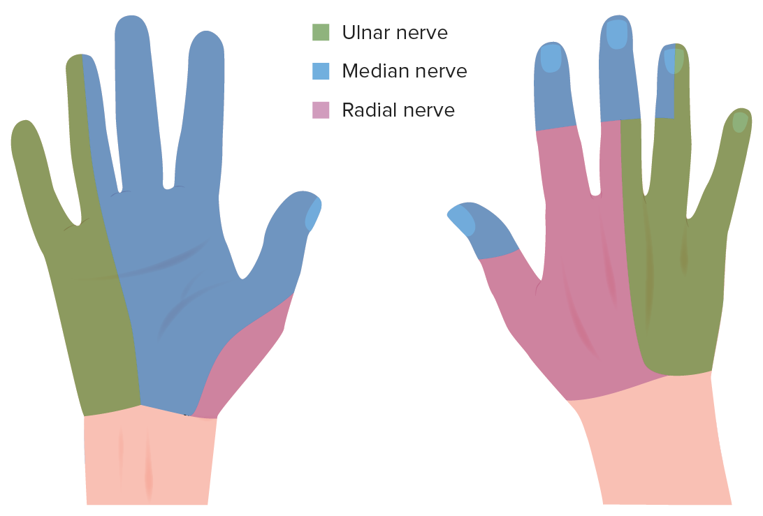
Which tendons are the origin of the lumbricals?
- Flexor digitorum profundus
- Flexor digitorum superficialis
- Palmaris longus
- Flexor digitorum indicis
- Flexor pollicis longus
Which statement regarding the lumbricals is correct?
- Lumbricals 1 and 2 originate from the lateral 2 tendons of the flexor digitorum profundus.
- Lumbricals 1 and 2 are bipennate.
- Lumbricals 3 and 4 are unipennate.
- Lumbricals 3 and 4 originate from the lateral 2 tendons of the flexor digitorum profundus.
- All lumbricals are unipennate.
Customer reviews
5,0 of 5 stars
| 5 Stars |
|
5 |
| 4 Stars |
|
0 |
| 3 Stars |
|
0 |
| 2 Stars |
|
0 |
| 1 Star |
|
0 |


